Human iPSC-Derived Cardiac Cells: i-HCm
Cryopreserved iPSC-Derived Cardiac Cells: Cardiomyocyte cell preparation derived from Human Induced Pluripotent Stem Cells
Size: 1 Cryovial
Price: $870.00
Description
Human iPSC-Derived Cardiac Cells (i-HCm) — alternatively written as hiPSC-derived cardiomyocytes, hiPSC-CMs, or referred to as induced pluripotent stem cell-derived cardiac muscle cells — are highly specialized cardiac myocytes generated through the directed differentiation of human induced pluripotent stem cells (iPS cells). Historically, the scarcity of viable human heart tissue has limited cardiovascular discovery and cardiotoxicity screening. By capturing a human genetic background in vitro, i-HCm serve as a crucial translational bridge that complements primary rodent models and heterologous expression systems. These cells form structurally synchronized monolayers that exhibit spontaneous electrical activity and contraction, serving as a robust platform for investigating human heart function and building models for conditions ranging from heart failure to complex inherited channelopathies. To maintain their specialized phenotype and support long-term cell survival, they are cultured using optimized, serum-free cardiac maintenance media.
While i-HCm are generated in culture dishes, their in vivo counterparts are the working myocytes found natively throughout the muscle layers of the human heart, particularly within the thick walls of the left ventricle. In a living patient, these cells work alongside adjacent vascular cells (such as endothelial cells and pericytes) to maintain blood flow and regulate systemic blood pressure. In clinical pathology — such as that studied by the American Heart Association — malfunctions across these tissue layers can manifest as chest pain, myocardial infarction, and full cardiac arrest. While i-HCm model critical cellular contributors to these conditions, investigators must note that single-cell cultures cannot recapitulate whole-organ hemodynamics or the complex, macro-scale physiology of a living heart. By pulling from diverse donor cohorts, investigators can study how variations in patient genomes alter myocyte physiology, comparing in vitro readouts against clinical diagnostics like a cardiac mri or patient histories of atrial fibrillation.
In laboratory settings, primary i-HCm provide an indispensable human-background platform for exploring genetic heart diseases, validating therapies for congestive heart failure, and conducting high-throughput safety pharmacology. Numerous studies published in journals like Cel Rep and J Cell Mol Med utilize patient-derived i-HCm to model the mechanobiology seen in obstructive hcm patients. By collaborating with specialized hypertrophic cardiomyopathy center investigators, researchers use these lines to screen small-molecule inhibitors designed to reduce hyper-contractility and alleviate the severe outflow obstructions characteristic of obstructive hypertrophic cardiomyopathy.
Because unanticipated cardiotoxicity remains a principal driver of late-stage drug failures, safety pharmacologists integrate i-HCm into multi-component paradigms like the Comprehensive In Vitro Proarrhythmia Assay (CiPA) initiative. Within the CiPA framework, i-HCm evaluations serve as a key component for assessing proarrhythmic risk, operating alongside high-throughput ion-channel data and in silico computational modeling rather than acting as the sole metric. Utilizing high-density Multi-Electrode Array (MEA) analysis and optical mapping, researchers screen novel chemical entities for drug-induced QT interval prolongation or structural breakthroughs, driving advancements in safety screening and cardiac regeneration.
Human iPSC-Derived Cardiac Cells contain i-HCm make a powerful in vitro platform to study cardiac physiology and disease, and to assess therapeutic compound efficacy, safety and toxicity1-3.
These iPSC-Derived Cardiac Cells are differentiated from integration-free Human Induced Pluripotent Stem Cells (HiPSC), which are validated for pluripotency and cultured under feeder-free conditions before cryopreservation. Through a robust serum-free, chemically defined protocol, high yields of fully functional Cardiac Cells are obtained and cryopreserved. Of note, the i-HCm and other cardiac cell populations are not purified through genetic selection, and thus there is no risk of genotoxic stress due to molecular manipulation. Furthermore, cellular preparations include accessory cellular populations resident in the human heart, which are crucial for correct cardiomyocyte physiology2, 4.
Characterization
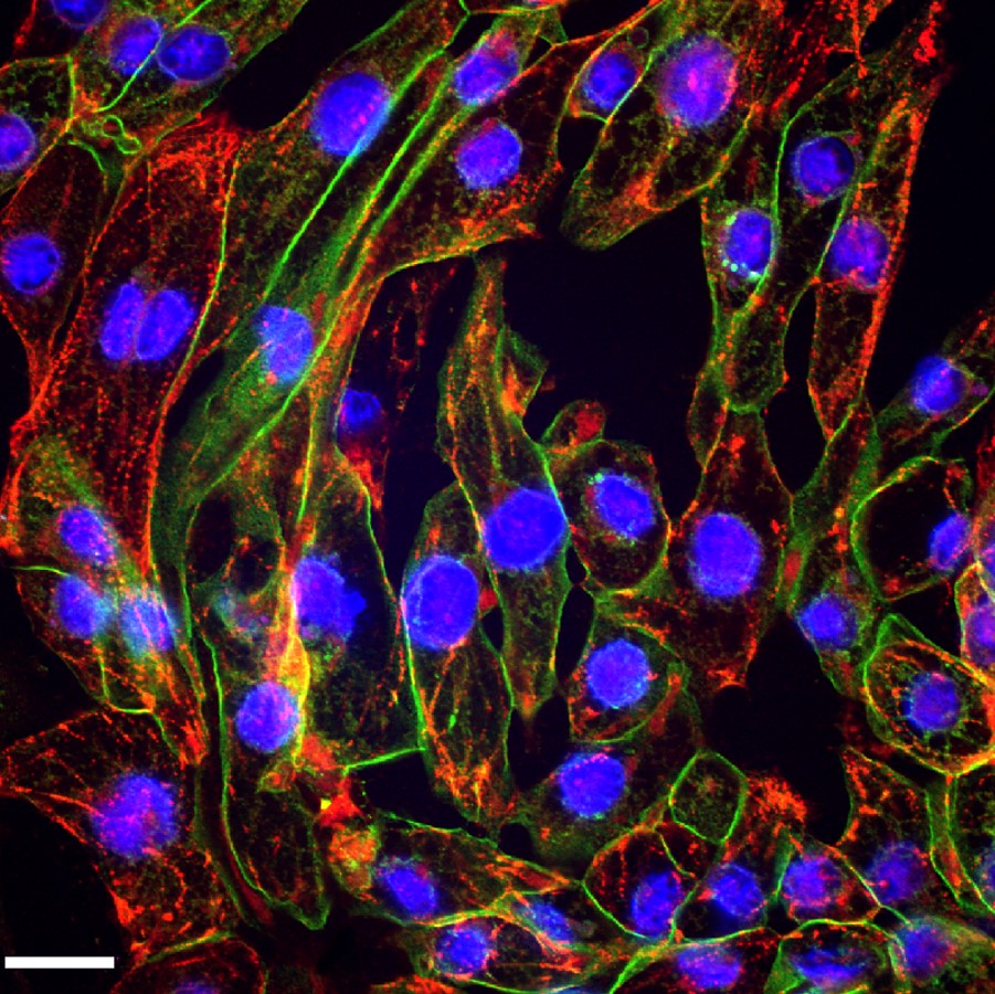
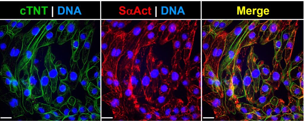
Post-thawing viability and plating efficiencies of the cells are typically higher than 50%. After plating at the recommended density, the cells will show spontaneous synchronized beating, and will express typical cardiomyocyte markers, such as the contractile proteins cardiac troponin T (cTNT) and sarcomeric alpha actinin (SaAct). The cardiomyocyte content of our preparations is determined by flow cytometry analysis and quantification of cardiac troponin T positive cells. These iPSC-Derived Cardiac Cells are also tested to ensure the absence of microorganism contaminants.
Applications
- Independent assays: From electrophysiology and multi-electrode array analysis to high content microscopy and viability screens.
- Cardiac Disease modeling: To date, interesting phenotypes of several genetic-based cardiomyopathies have been successfully replicated in a dish through the use of patient-specific i-HCm. Examples include Familial Hypertrophic, Dilated, and Arrhythmogenic Right Ventricular Cardiomyopathies1-3.
- Cardiac safety and toxicity: Cardiac toxicity is an important contributor for the failure of therapeutic agents in late stages of clinical trials, as well as for the removal of approved drugs from the market. Through the CiPA initiative, which brings together researchers from institutions such as the FDA, academic institutions and pharmaceutical companies, i-HCm are being evaluated as an integral tool for the safety assessment of developing drugs5,6.
References
- Zanella F., R. Lyon and F. Sheikh. 2014. Modeling Heart Disease in a Dish: From Somatic Cells to Disease-Relevant Cardiomyocytes. Trends Cardiovasc Med, 24:32-44.
- Passier R., V. Orlova and C. Mummery C. 2016. Complex Tissue and Disease Modeling using hiPSCs. Cell Stem Cell, 19:309-321.
- Moretti A., K. Laugwitz, T. Dorn, D. Sinnecker and C. Mummery. 2013. Pluripotent stem cell models of human heart disease. Cold Spring Harb Perspect Med. 2013; 3(11).
- Burridge P., S. Metzler, K. Nakayama, O. Abilez, C. Simmons, M. Bruce, Y. Matsuura, P. Kim, J. Wu, M. Butte, F. Huang and P. Yang. 2014. Multi-cellular interactions sustain long-term contractility of human pluripotent stem cell-derived cardiomyocytes. Am J Transl Res, 22:724-735.
- Cavero, I. and H. Holzgrefe. 2014. Comprehensive in vitro Proarrhythmia Assay, a novel in vitro/in silico paradigm to detect ventricular proarrhythmic liability: a visionary 21st century initiative. Expert Opin Drug Safety, 13:745-758.
- Fermini B., J. Hancox, N. Abi-Gerges, M. Bridgland-Taylor, K. Chaudhary, T. Colatsky, K. Correll, W. Crumb, B. Damiano, G. Erdemli, G. Gintant, J. Imredy, J. Koerner, J. Kramer, P. Levesque, Z. Li, A. Lindqvist, C. Obejero-Paz, D. Rampe, K. Sawada, D. Strauss and J. Vandenberg. 2016. A New Perspective in the Field of Cardiac Safety Testing through the Comprehensive In Vitro Proarrhythmia Assay Paradigm. J Biomol Screen, 21:1-11.
Details
| Source | Derived from Human iPSC of a single donor | |
|---|---|---|
| QC | No bacteria, yeast, fungi, mycoplasma, virus | |
| Bioassay | Spontaneous synchronized beating | |
| Character | cTNT and SαAct positive | |
| CofA | Donor, gender, tissue source, disease status, cell viability (post-thaw), passage #, karyotype, immunostaining markers | |
| Cryovial | Non-proliferative 2,000,000 i-HCm in freezing med. | |
| Medium | Cells manufactured, maintained in serum-free, feeder-free culture conditions | |
| Plating Efficiency | ≥ 50% | |
| Seeding Density | 250,000 cells/cm2 | |
| Storage | Liquid Nitrogen | |
| Applications | Non-commercial research use only (RUO), not for human or clinical use |
Human Cardiomyocyte Differentiation Medium Kit* (017DK)
| Cat. No. | Medium | Vol | Store |
|---|---|---|---|
| 017D1-30 | Differentiation 1 | 30ml | 4°C |
| 017D2-30 | Differentiation 2 | 30ml | 4°C |
| 017D3-30 | Differentiation 3 | 30ml | 4°C |
| 019-50 | Selection | 50ml | 4°C |
| 021XF-30 | Maintenance** | 30ml | 4°C |
Resources
FAQs
Need More Help?
Visit our comprehensive FAQ page for detailed answers to common questions.
Need More Help?
Visit our comprehensive FAQ page for detailed answers to common questions.
Primary Cell FAQs Powered by Bioz