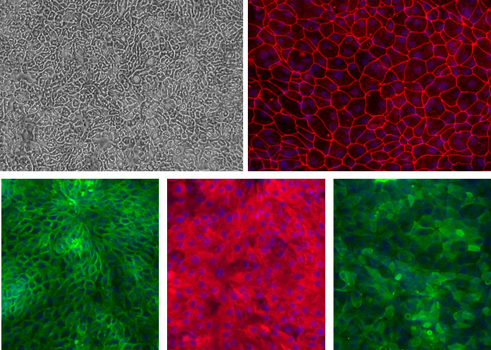
Human Jejunum Epithelial Cells (HJeEpC) and other Adult GI EpC monolayers. Top left: Phase contrast of Ileum EpC. Top right (Red): ZO-1 of Duedenum EpC. Bottom left (Green): Na+/K+ ATPase of Colon EpC. Bottom middle (Red): pan-Cytokeratin of Gastric EpC. Bottom right (Green) Villin of Jejunum EpC. Blue: DAPI.
Human Jejunum Epithelial Cells: HJeEpC
Human Jejunum Epithelial Cells (HJeEpC) are a specialized population of epithelial cells derived from the jejunum, the middle section of the small intestine.
Description
Human Jejunum Epithelial Cells (HJeEpC) — sometimes referred to in literature under the broader term human intestinal epithelial cells (HIEC) — are a specialized population of cells derived from the jejunum, the middle section of the small intestine. As the primary site for nutrient absorption, the jejunum plays a vital role in human metabolism and homeostasis. These cells line the intestinal lumen, forming a highly organized, single-layered epithelium that balances the uptake of essential nutrients from food with the maintenance of a protective barrier against luminal pathogens and toxins.
The intestinal epithelium is organized into a dynamic structure of crypts and villi, representing among the most rapidly self-renewing tissues in the adult human body. From birth to adulthood, the intestinal lining maintains this high turnover rate through a continuous cycle of proliferation and terminal differentiation. Within the jejunal epithelium, cells fulfill specific roles:
- Enterocytes: The most abundant cell type, specialized for the transport of nutrients and expressing significant brush-border (membrane-associated) digestive enzymes, such as disaccharidases and peptidases, to facilitate the final steps of digestion.
- Goblet Cells: Specialized cells interspersed among enterocytes that secrete protective mucus, forming a critical component of the mucosal defense system.
- Structural Specializations: To maximize surface area for uptake, the cells feature microvilli—tiny epithelial protrusions that significantly enhance the efficiency of solute and water transport.
The main function of the crypt-villus axis is to ensure the constant renewal of the epithelium. Transit-amplifying cells originate in the crypts, where they undergo multiple rounds of division before differentiating into specialized epithelial lineages. As they mature and migrate toward the tip of the villus, they are shed into the gut lumen, a process critical for tissue homeostasis.
The intestinal barrier is not defined by tight junctions alone; it is a complex, multifaceted defense system. It integrates the physical barrier provided by tight junctions, the protective mucus layer, the secretion of antimicrobial factors, innate immune signaling, and the high rate of epithelial turnover to effectively protect the underlying tissues from luminal threats.
HJeEpC provide an outstanding in vitro model for investigating the physiology of the gastrointestinal (GI) tract. They are particularly valuable as a place to study:
- Disease Modeling: Research into Inflammatory Bowel Disease (IBD), including Crohn’s disease and ulcerative colitis, as well as intestinal cancers.
- Drug Discovery: Serving as a reliable test platform for evaluating potential drug candidates, disease modulators, and toxicological responses.
- Barrier Function: Because HJeEpC form a polarized monolayer with well-developed intercellular junctions — confirmed by markers such as Villin (epithelial identity), ZO-1 (tight junction integrity), and Na+/K+ ATPase (polarity) — they are ideal for functional analyses of epithelial integrity and permeability.
HJeEpC are essential for understanding the complexities of intestinal health and disease. By modeling the physiological environment of the jejunum in vitro, researchers can explore the mechanics of nutrient uptake and multi-layered barrier defense, fostering advancements in regenerative therapy, drug screening, and the clinical management of chronic GI disorders.
The intestinal epithelium is a single layer of cells organized into crypts and villi, known as the most rapidly self-renewing tissue in adult mammals. The cells that line the intestinal lumen perform the primary functions of digestion, water and nutrient absorption, and forms a barrier against luminal pathogens.
Transit-amplifying cells spend approximately two days in the intestinal crypts, dividing 4–5 times before terminally differentiating into specialized intestinal epithelial cell types. In the small intestine, the surface area is dramatically enlarged through epithelial protrusions called villi. Three days after their terminal differentiation, the cells reach the tip of the villus, undergo spontaneous apoptosis, and are shed into the gut lumen.
CAI’s intestinal epithelial culture system provides outstanding resource for investigation of intestinal epithelial cell physiology related to GI infection, inflammatory bowel disease (IBD) like Crohn’s disease, ulcerative colitis, and intestinal cancer. Our epithelial cell culture system can be efficiently used as a test platform for the potential drug candidates and disease modulators. Other applications of this culture system include functional analysis of intestinal epithelium, GI disease modeling, and regenerative therapy preclinical testing such as drug compound screening and other validation assays.
With optimized, defined culture media from CAI, the Intestinal Epithelial Cells can be seeded and maintained for as long as 8 days. Epithelial Cells grown in CAI medium form a monolayer of polarized epithelial cells with tight junction formation as evidenced by Villin (apical marker), Na+/K+ ATPase (basolateral marker), ZO-1 (tight junction marker) and pan-Cytokeratin (epithelial marker) staining.
Details
| Tissue | Normal healthy human intestine. | |
|---|---|---|
| QC | No bacteria, yeast, fungi, mycoplasma, virus. | |
| Bioassay | Attach, spread, in Culture Medium. | |
| Cryovial | 500,000 HJeEpC in Freezing Medium. | |
| Kit | Cryovial frozen HJeEpC (732Je-05a), Culture Med (716DC-50), Coating Solution (1024-05), Thawing Solution (716T-20). | |
| Cultured | Shipped in flasks or plates in medium. | |
| Doublings | Cells do not divide and cannot be passaged. | |
| Applications | Laboratory research use only (RUO). Not for human, clinical, diagnostic or veterinary use. |
Resources
FAQs
Need More Help?
Visit our comprehensive FAQ page for detailed answers to common questions.
Need More Help?
Visit our comprehensive FAQ page for detailed answers to common questions.
Primary Cell FAQs