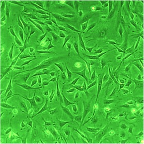
Rat Cardiac Fibroblasts: RCF. Isolated from rodent heart tissue.
Rat Cardiac Fibroblasts: RCF
Rat Cardiac Fibroblasts (HCF) are fibroblast cells from the rodent heart.
Description
Rat Cardiac Fibroblasts (RCF) are the most prevalent cell type in the rodent heart, comprising approximately 60–70% of the total cardiac cell population. These rat primary cells are indispensable for maintaining the integrity of the cardiac matrix through constant extracellular matrix synthesis and turnover. Because they are central to both structural support and the mechanical signaling of the myocardium, RCF provide an excellent model system for studying cardiac fibroblast biology, including the regulation of cell growth, survival, inflammation, hormone secretion, and metabolism. To ensure consistent experimental results, high quality RCF lots are rigorously screened to confirm they are free from contaminants such as mycoplasma or fungi, ensuring that researchers are working with healthy, live cells that accurately reflect the native extracellular matrix environment.
RCF are primary drivers of cardiac fibrosis and structural remodeling following various pathological conditions:
- Activation and Differentiation: Upon cardiac injury — such as that experienced during myocardial infarction or ischemia-reperfusion injury — these cells undergo phenotypic activation into the cardiac myofibroblast state. This transition is characterized by enhanced contractile machinery, altered integrin expression, and the secretion of pro-fibrotic factors that contribute to maladaptive scar formation.
- Signaling and Crosstalk: These fibroblasts communicate with surrounding cardiac myocytes and endothelial cells through the secretion of paracrine growth factors and cytokines. Signaling cascades, including those mediated by the PDGF receptor, regulate this transition. Investigating these signaling pathways is vital for understanding how the heart responds to pathological hypertension stressors and progresses toward cardiac hypertrophy and, eventually, heart failure.
The versatility of RCF allows investigators to model the complex microenvironments of the heart with high fidelity:
- Comparative Molecular Biology: Researchers frequently utilize RCF alongside models of the mouse heart to delineate conserved mechanisms of injury and repair. By characterizing changes in gene expression and protein secretion during transitions from healthy to diseased states, these fibroblasts serve as a baseline for understanding how the heart compensates for stress.
- Experimental Precision: Through techniques such as flow cytometry, researchers can precisely monitor cellular states, ensuring that their models accurately reflect the heterogeneity of the cardiac tissue. Because these fibroblasts orchestrate the environment for other cells, they are frequently utilized in co-culture systems — often involving mouse endothelial cells — to simulate the complex interactions that maintain heart rhythm and contractile function.
Because RCF play a dominant role in the organ’s response to damage, they are a primary target for therapeutic intervention:
- Regenerative Medicine: Beyond their structural role, studies into these fibroblasts help researchers identify ways to modulate cardiac fibrosis without hindering essential wound-healing processes. While other lineages, such as stem cells, are explored for their potential to replace damaged tissue, the native fibroblast population remains the primary gatekeeper of the structural landscape.
- Broad Applications: By providing a reliable platform for high-throughput screening, RCF enable the identification of compounds that can inhibit maladaptive activation or promote a more regenerative response, making them a cornerstone for any laboratory investigating the molecular basis of cardiovascular disease.
Rat Cardiac Fibroblasts (RCF) are the most prevalent cell type in the rodent’s heart, making up 60-70 % of all cells. They are known to play key roles in the development of cardiac disease and hypertrophy. Rat Cardiac Fibroblasts from Cell Applications, Inc. provide an excellent model system to study many aspects of heart function and pathophysiology, including the regulation of cell growth, survival, inflammation, hormone secretion and metabolism.
Details
| Tissue | Normal healthy rat heart | |
|---|---|---|
| QC | No bacteria, yeast, fungi, mycoplasma | |
| Bioassay | Attach, spread, proliferate in Growth Med | |
| Cryovial | 500,000 RCF (2nd passage) in frozen in Basal Medium w/ 10% FBS, 10% DMSO | |
| Kit | Cryovial frozen RCF (R306-05a/-05n), Gr Med (R116-500), Subcltr Rgnt Kit (090K) | |
| Proliferating | Shipped within 3 days after plating in Cltr Medium (flasks or plates) | |
| Doublings | At least 8 | |
| Applications | Laboratory research use only (RUO). Not for human, clinical, diagnostic or veterinary use. |
Resources
FAQs
Need More Help?
Visit our comprehensive FAQ page for detailed answers to common questions.
Need More Help?
Visit our comprehensive FAQ page for detailed answers to common questions.
Primary Cell FAQs Powered by Bioz