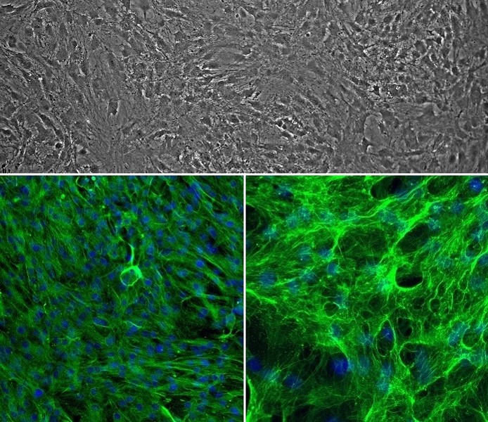
Rat Perineurial Fibroblasts (RPF) revived (top) and stained for Vimentin (green, bottom left), Fibronectin (green, bottom right), and DAPI (blue).
Rat Perineurial Fibroblasts: RPF
Rat Perineurial Fibroblasts (RPF) are primary cells of mesenchymal origin that form the perineurium, one of the three connective tissue compartments of the peripheral nerve.
Description
News Release
Rat Perineurial Fibroblasts (RPF) are primary cells of mesenchymal origin that form the perineurium, one of the three connective tissue compartments of the peripheral nerve. In vivo, the perineurium wraps around individual nerve fascicles, where it contributes to the perineurial barrier, limits diffusion, and supports endoneurial homeostasis while helping protect nerve bundles and modulate external stretching forces.
In experimental research, RPF provide an in vitro model system that can be used to study cellular and molecular processes relevant to peripheral nerve compartments. When cultured in fibroblast growth medium, proliferating rat perineurial fibroblasts can be utilized alongside other rat primary cells — such as rat dermal fibroblasts, rat pulmonary fibroblasts, endothelial cells, blood borne cells, and Schwann cells — to investigate mechanisms relevant to peripheral nerve injury, nerve gaps, nerve regeneration, and nerve repair. While ordinary fibroblasts and perineurial cells share core structural properties, perineurial fibroblasts can exhibit distinct ultrastructural features, such as pinocytotic vesicles, and contribute uniquely as a specialized cell type to peripheral nerve tissue architecture.
At the molecular level, Rat Perineurial Fibroblasts express the cytoplasmic protein vimentin, a Type III intermediate filament that forms part of the cytoskeleton. Vimentin filaments help maintain cell shape, cytoplasmic integrity, and cytoskeletal interactions, functioning in cell adhesion, migration, and intracellular signaling. In addition, RPF express fibronectin, a multifunctional extracellular matrix glycoprotein that plays major roles in cell morphology, adhesion, growth, migration, and extracellular matrix organization, and may be associated with perineurial-associated marker programs (e.g., epithelial membrane antigen or EMA), depending on culture conditions and phenotype. These molecular features and gene expression profiles support investigations into cellular responses following peripheral nerve trauma.
Rat Perineurial Fibroblasts (RPF) from Cell Applications, Inc. are of mesenchymal origin and form the perineurium, one of the three connective tissue compartments of the peripheral nerve. The perineurium wraps around nerve fascicles to form a metabolically active diffusion barrier. It provides protection to the nerve bundles, maintains the homeostasis of the endoneurium, modulates external stretching forces that exerts to the nerve, and regulates endoneurial pressures.
RPF and other cells of mesenchymal origin express the cytoplasmic protein Vimentin, a Type III intermediate filament that forms part of the cytoskeleton. By helping to maintain cell shape, cytoplasmic integrity and cytoskeletal interactions, Vimentin functions in cell adhesion, migration and signaling.
Perineurial fibroblasts also express fibronectin, a multifunctional extracellular matrix glycoprotein. Like vimentin, fibronectin plays major roles in cell morphology, cytoskeletal organization, adhesion, growth and migration. Fibronectin, which binds other ECM molecules, also functions in cell differentiation, embryonic development, hemostasis, phagocytosis, wound healing and repair.
Details
| Tissue | Normal healthy rat peripheral nerves | |
|---|---|---|
| QC | No bacteria, yeast, fungi, mycoplasma | |
| Character | Positive for Fibronectin | |
| Bioassay | Attach, spread, proliferate in Growth Med | |
| Cryovial | 500,000 RPF (1st passage) frozen in Basal Medium w/ 10% FBS, 10% DMSO | |
| Kit | Cryovial frozen RPF (R852-05n), Growth Medium (R827-500), Subcltr Rgnt Kit (090K) | |
| Proliferating | Shipped in Gr Med, 1st psg (flasks or plates) | |
| Doublings | At least 12 | |
| Applications | Laboratory research use only (RUO). Not for human, clinical, diagnostic or veterinary use. |
Resources
FAQs
Need More Help?
Visit our comprehensive FAQ page for detailed answers to common questions.
Need More Help?
Visit our comprehensive FAQ page for detailed answers to common questions.
Primary Cell FAQs