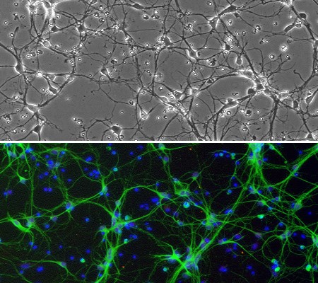
Cryopreserved Rat Striatal Neurons (RStN) revived and cultured for 8 days (top), stained (bottom) for β III-Tubulin (green), GFAP (red), and DAPI (blue).
Rat Striatal Neurons: RStN
Rat Striatal Neurons (RStN) are primary neural cells derived from the striatum of normal embryonic rats.
Description
Rat Striatal Neurons (RStN) are primary neural cells derived from the striatum of normal embryonic rats (typically isolated on embryonic day 18) using standardized methods. When cultured with rat neuron culture medium under recommended conditions, these cells rapidly arborize and form complex neurite networks within one week.
In vivo, the striatum is a subcortical structure within the forebrain that receives and processes input signals from the cerebral cortex and dopaminergic projections from the substantia nigra. Featuring robust interactions among cholinergic, dopaminergic, and GABAergic neurotransmitter systems, the striatum plays a central role in regulating voluntary movement, reward-based habit learning, addiction, procedural memory formation, and the neural mechanisms underlying cognitive dysfunctions and motor deficits in Parkinson’s disease.
In neurobiology research, Rat Striatal Neurons (RStN) provide a valuable in vitro model to study striatal-related physiological functions, pathological processes, and neuropharmacological responses. Cultures of striatal neurons can be applied to a wide array of experimental techniques, including toxicity testing, immunostaining, live cell imaging, co-culturing, and electrophysiology. Investigators typically characterize these cultures by confirming positive expression for neuronal markers such as βIII-tubulin (TUBB3), which is used to confirm neuronal identity and maturation before functional testing.
Rat Striatal Neurons (RStN) are derived from striatum of normal embryonic rat (gestation day 18) by standardized methods. When cultured under the recommended conditions, RStN arborize and form complex neurite network in one week. RStN Stain positive for β III-Tubulin.
The striatum is a subcortical part of the forebrain. It receives and processes input signals from different parts of the cerebral cortex. With its robust interactions among cholinergic, dopaminergic, and GABAergic neurotransmitter systems, striatum plays a key role in voluntary movement, reward-based habit learning, addiction, procedural memory formation, and cognitive dysfunctions in Parkinson’s disease. Studies using striatal neurons will allow a better understanding of the striatal-related functions and diseases. Cultures of striatal neurons can be applied for a variety of experiments including toxicity test, immunostaining, live cell imaging, co-culturing, electrophysiology, and more.
Details
| Tissue | Normal healthy rat striatum | |
|---|---|---|
| QC | No bacteria, yeast, fungi, mycoplasma | |
| Character | Positive for β-III Tubulin | |
| Bioassay | Plate on Poly-D-Lysine coated surface, Arborize to form neurite network in Culture Med | |
| Cryovial | 1M RStN in Serum-free Frzng Med (042-50) | |
| Kit | Cryovial frzn RStN (R8812N-10), Neuron Ctng Soln I (027-05), Plating (R817P-10) & Cultr Med (R817-100) | |
| Doublings | N/A – Neurons don’t proliferate in vitro | |
| Applications | Laboratory research use only (RUO). Not for human, clinical, diagnostic or veterinary use. |
Resources
FAQs
Need More Help?
Visit our comprehensive FAQ page for detailed answers to common questions.
Need More Help?
Visit our comprehensive FAQ page for detailed answers to common questions.
Primary Cell FAQs Powered by Bioz