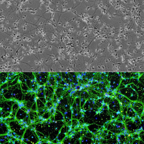
Cryopreserved Rat Spinal Cord Neurons (RSpN) revived and cultured for 6 days (top), stained (bottom) on day 7 for β III-Tubulin (green), GFAP (red), and DAPI (blue).
Rat Spinal Cord Neurons: RSpN
Cryopreserved Rat Spinal Neurons (RSpN): Frozen RSpN (2×10^6)
Size: 1 Cryovial
Price: $743.00
Description
Rat Spinal Cord Neurons (RSpN) are primary cells isolated from the embryonic spinal cord of normal Sprague-Dawley rats (typically gestation day 15) using standardized methods. Upon isolation and cultivation under recommended conditions, RSpN rapidly arborize, forming complex, interconnected neurite networks within one week. Characterized by their positive expression of β III-tubulin, these nerve cells serve as a critical in vitro model for studying the intricate physiology of the central nervous system. As the primary conduit for transmitting neural signals between the brain and the body, the spinal cord is essential for somatosensory and motor functions; RSpN allow researchers to bridge the gap between basic developmental biology and the clinical challenges posed by spinal cord injury and degenerative pathology.
The spinal cord is a complex environment consisting of nervous tissue, glial support cells, and a dense network of axons and nerve fiber tracts. In the context of cns injury, this architecture is often disrupted, leading to the formation of restrictive scar tissue and the loss of functional connections.
- Mechanistic Research: RSpN are indispensable for investigating the physiopathology of conditions such as Amyotrophic Lateral Sclerosis (ALS), Friedreich’s ataxia, and spinal muscular atrophies. By observing the interplay between host neurons and their environment, scientists can better understand how acute spinal cord injury or chronic spinal cord contusion leads to the progressive loss of motor function.
- Regenerative Strategies: A primary focus of modern neurobiology is promoting regeneration within the spinal injuries microenvironment. Researchers utilize RSpN to evaluate bioactive materials and therapeutic scaffolds designed to bypass a lesion site. These experiments often examine how transplanted neural progenitor cells or neural stem cells can integrate into the existing network to facilitate functional recovery.
Cultures of RSpN support a diverse range of experimental applications, from basic neurotoxicity screenings to complex circuit analysis.
1. Advanced Experimental Platforms
RSpN are uniquely suited for:
- Electrophysiology and Imaging: Real-time live cell imaging and patch-clamp electrophysiology allow investigators to track brain activity and neuronal signaling patterns that are often obscured in in vivo models.
- Co-culture Systems: By incorporating astrocyte layers or blood vessels, these cultures recreate the tripartite synapse (neuron-astrocyte-microglia interaction) and the neurovascular unit, providing a more accurate representation of the adult rat spinal cord microenvironment.
2. Comparative and Translational Context
While RSpN provide the foundation for understanding spinal circuitry at the thoracic level and beyond, they are frequently utilized alongside other models. Comparative studies often evaluate RSpN against human embryonic stem cells or neurons derived from the rat brain to validate potential therapies for neurodegenerative disease. Furthermore, investigating how RSpN respond to chronic pain pathways—or how they are influenced by different brain region inputs—is essential for refining the next generation of treatments for human patients.
As researchers continue to optimize the advanced features of these culture models, RSpN remain a cornerstone of the rat model and animal study frameworks, offering a clear window into the delicate balance of neural health and the devastating consequences of trauma.
Rat Spinal Cord Neurons (RSpN) are derived from spinal cords of normal embryonic rat (gestation day 15) by standardized methods. When cultured under the recommended conditions, RSpN arborize and form complex neurite network in one week. RSpN Stain positive for β III-Tubulin.
The spinal cord is consisted of tubular bundles of nervous tissues and support cells. Extending from the medulla oblongata of the brainstem, the spinal cord transmits neural signals between the brain and the rest of the body, and plays a key role in somatosensory and motor functions. Spinal cord injuries, diseases and degeneration, such as Friedreich’s ataxia, Amyotrophic lateral sclerosis, and spinal muscular atrophies, cause weakening or loss of sensory and motor functions. Studies using spinal cord neurons will allow a better understanding of the disease mechanisms, physiopathologies, and advancement in drug development and therapies. Cultures of spinal cord neurons can be applied for a variety of experiments including toxicity test, immunostaining, live cell imaging, co-culturing, electrophysiology, and more.
Details
| Tissue | Normal healthy rat spinal cord | |
|---|---|---|
| QC | No bacteria, yeast, fungi, mycoplasma, virus | |
| Character | Positive for β-III Tubulin | |
| Bioassay | Plate on Poly-D-Lysine coated surface, Arborize to form neurite network in Culture Med | |
| Cryovial | 2 or 4M RSpN in Ser-Fr Frzng Med (042-50) | |
| Doublings | N/A – Neurons don’t proliferate in vitro | |
| Applications | Laboratory research use only (RUO). Not for human, clinical, diagnostic or veterinary use. |
Resources
FAQs
Need More Help?
Visit our comprehensive FAQ page for detailed answers to common questions.
Need More Help?
Visit our comprehensive FAQ page for detailed answers to common questions.
Primary Cell FAQs