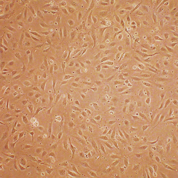
Human Aortic Smooth Muscle Cells: Type 2 Diabetes: HAOSMC-T2D
Human Aortic Smooth Muscle Cells: Type 2 Diabetes (HAOSMC-T2D) are specialized vascular smooth muscle cells isolated from the medial layer of the human aorta tissue of donors clinically diagnosed with type 2 diabetes.
Description
Human Aortic Smooth Muscle Cells: Type 2 Diabetes (HAOSMC-T2D) are specialized vascular smooth muscle cells isolated from the medial layer of the human aorta tissue of donors clinically diagnosed with type 2 diabetes. As a highly distinct, diseased cell type, HAOSMC-T2D exhibit structural and metabolic alterations in vitro compared to healthy smooth muscle counterparts. Characterized as primary cells rather than an immortalized cell line, they maintain a “diseased phenotype” imprint that reflects the chronic in vivo physiological stresses of metabolic dysfunction. However, the extent of phenotypic switching and the presentation of disease-associated traits depend heavily on donor variability, comorbidities, clinical medication history, ischemic time, and the in vitro passage number, meaning these alterations are not uniformly identical across all T2D-derived fractions. For definitive validation, characterization protocols verify that these diverse primary cells remain strongly positive for smooth muscle cell-specific alpha smooth muscle actin (also known as α-SMA) expression, confirming their functional identity.
In the human body, these smooth muscle cells form the primary structural and contractile component of the tunica media within the human aorta, the largest elastic artery in the cardiovascular system. In diabetic patients, these cells are chronically exposed to pathological, systemic microenvironments characterized by elevated blood glucose levels and persistent pro-inflammatory signaling. To capture this specific disease state for research, the cells are isolated directly from the aortas of deceased or surgical donors with documented type 2 diabetes. Within this clinical context, studying these cells helps investigators understand structural wall failures and macrovascular disease progression, which can contribute to risk factors associated with aortic disease, including conditions that may predispose to dissection.
In vivo, the principal function of healthy vascular smooth muscle cells is to regulate arterial blood pressure and tissue perfusion through coordinated contraction and relaxation, while synthesizing the extracellular matrix (ECM) necessary for vascular elasticity. In a chronic diabetic state, these cells can undergo a phenotypic shift from a contractile state to a more synthetic, proliferative state. This phenotypic switching can alter normal cell function, leading to accelerated cell proliferation, migration into the intima, and dysregulated cell death pathways. While VSMC dysfunction is a major factor, it acts alongside ECM degradation, local inflammation, and altered hemodynamics to contribute to pathogenic vascular remodeling, which in turn plays a role in the severe macrovascular complications and accelerated atherosclerosis typically observed in individuals with type 2 diabetes.
In laboratory settings, HAOSMC-T2D are utilized in specialized cell culture systems to investigate the molecular mechanisms linking metabolic disorders to aggressive vascular disease. When maintained in an optimized growth medium, researchers manipulate ambient glucose concentrations to study how hyperglycemia dictates altered gene expression and modifies specific cytokines and growth factor signaling pathways. Because vascular pathologies involve intricate cellular cross-talk, these cells are frequently co-cultured with aortic endothelial cells to analyze cell-to-cell signaling, restenosis risks, and mechanisms of angiogenesis in vitro during tissue repair. By tracing altered genes and testing advanced drug or gene delivery systems on these diseased cells, investigators leverage HAOSMC-T2D as a translationally relevant in vitro model to develop novel therapeutic interventions, optimize endovascular stent coatings, and mitigate occlusive vasculopathies in diabetic patient populations.
- Cytokines and growth factor signaling pathways implicated in the molecular regulation of smooth muscle cell proliferation, migration, and overall vascular function
- Hyperglycemia-related risk factors for atherosclerosis in diabetes patients, as well as effects of ethanol on vascular calcification
- ECM deposition and its role in cardiovascular health, repair of damaged vasculature and successful tissue engineering
- Mechanisms and effects of mechanoregulation on proliferation and function of smooth muscle cells
- Advanced stent technology, including novel surface materials
- Drug and gene delivery systems
- Restenosis and other occlusive vasculopathies
Characterization: Positive for smooth muscle cell specific alpha-actin expression. Type 2 Diabetes (T2D) Cellular Disease Models For T2D research and drug discovery, Cell Applications offers multiple cell types, isolated from donors where genetics and lifestyle factors contributed to insulin resistance and high blood sugar levels.
Details
| Tissue | Human aorta from donor with Type 2 Diabetes (T2D) | |
|---|---|---|
| QC | No bacteria, yeast, fungi, mycoplasma, virus | |
| Character | Smooth muscle specific α-actin positive | |
| Bioassay | Attach, spread, proliferate in Growth Med | |
| Cryovial | 500,000 HAOSMC (2nd passage) frozen in Basal Medium w/ 10% FBS, 10% DMSO | |
| Kit | Croyvial frozen HAOSMC-T2D (354T2D-05a), Growth Medium (311-500), Subcltr Rgnt Kit (090K) | |
| Proliferating | Shipped in Gr Med, 3rd psg (flasks or plates) | |
| Doublings | At least 16 | |
| Applications | Laboratory research use only (RUO). Not for human, clinical, diagnostic or veterinary use. |
FAQs
Need More Help?
Visit our comprehensive FAQ page for detailed answers to common questions.
Need More Help?
Visit our comprehensive FAQ page for detailed answers to common questions.
Primary Cell FAQs