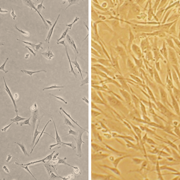
Human Coronary Artery Smooth Muscle Cells: Asthma: HCASMC-AS
Human Coronary Artery Smooth Muscle Cells: Asthma: HCASMC-AS
Human Coronary Artery Smooth Muscle Cells from Asthma Donors (HCASMC-AS) are primary human smooth muscle cells isolated from the coronary arteries of individuals diagnosed with asthma.
Description
Human Coronary Artery Smooth Muscle Cells from Asthma Donors (HCASMC-AS) are primary human smooth muscle cells isolated from the coronary arteries of individuals diagnosed with asthma. While researchers frequently utilize standard airway smooth muscle (ASM) or bronchial smooth muscle models to study localized respiratory diseases, HCASMC-AS serve as a distinct macrovascular model.
In an in vitro cell culture environment, these specialized mesenchymal cells allow investigators to explore systemic, vascular alterations. These cells provide a crucial tool for determining how systemic inflammatory environments linked to chronic respiratory diseases correlate with macrovascular smooth muscle behavior.
In vivo, these cells populate the tunica media of the coronary arteries, providing the structural integrity and vasomotion necessary to regulate cardiac blood flow. This vascular niche is fundamentally distinct from the respiratory system, where airway smooth muscle cells (or asm cell populations) encircle the airway wall.
In the respiratory tract of asthmatic patients, chronic inflammation directly insults the airway epithelium, causing the underlying connective tissue and laminae to undergo profound airway remodeling. This remodeling narrows the airway lumen and increases overall asm mass in asthma patients.
The biological link between these two distinct physiological compartments—the asthmatic airway and the coronary vasculature—remains an active area of intensive scientific inquiry. While large-scale epidemiological data consistently demonstrates that severe asthma correlates with an elevated risk of ischemic heart disease and acute myocardial infarction, the exact underlying mechanisms are still being elucidated. A primary working hypothesis is that chronic inflammatory mediators originating within asthmatic airways may enter systemic circulation, exposing distant coronary smooth muscle to a prolonged, low-grade inflammatory state.
The baseline function of coronary smooth muscle cells is to contract and relax dynamically to manage myocardial perfusion. However, chronic exposure to circulating inflammatory mediators is thought to potentially drive pathological smooth muscle remodeling, shifting these cells from a quiescent state to an active state.
1. Phenotypic Switching and Proliferation
Under inflammatory stress, coronary smooth muscle cells can undergo a phenotypic switch, losing their contractile markers—such as alpha smooth muscle actin (α-SMA)—and transitioning into a synthetic phenotype. This switch can accelerate cell proliferation (specifically smooth muscle cell proliferation), mimicking the hyperproliferative behavior observed in asthmatic asm cells during respiratory asm proliferation.As illustrated above, this enhanced asm cell proliferation and migration into the intima contributes to neointimal hyperplasia, altering how the cells deposit extracellular matrix proteins and remodel vascular plaques.
2. Hyper-Reactivity Pathways
At the cellular level, inflammatory signaling can lower the threshold for vascular spasm—a phenomenon reflected clinically in Kounis syndrome (allergic vasospasm), where acute mast cell degranulation triggers coronary constriction. Some studies report altered signaling cascades in inflamed vascular smooth muscle cells, giving researchers a molecular blueprint to test in HCASMC-AS to determine if a lower threshold for hyper-contraction persists in vitro.
HCASMC-AS serve as an essential model to bridge the gap between pulmonary inflammation and macrovascular disease, helping to unravel a highly complex systemic axis.
Investigating Systemic Biomarkers
Researchers utilize HCASMC-AS to test how specific cytokines drive or sustain vascular wall activation. For example, in an in vitro assay, scientists can apply a neutralizing antibody to the culture media to block IL-6 signaling pathways. This helps clarify whether coronary smooth muscle alterations are driven by continuous local exposure to specific cytokines, or if they represent independent phenotypic shifts separate from surrounding immune cells and inflammatory cells.
Stem Cell and Regenerative Studies
By evaluating how primary HCASMC-AS behave compared to healthy controls, scientists can identify specific microenvironmental cues that guide vascular disease. These findings inform stem cell research and tissue engineering, helping investigators design vascular grafts and targeted therapies that resist the pro-inflammatory, hyperproliferative signaling typically found in patients with chronic inflammatory diseases.
Human Coronary Artery Smooth Muscle Cells (HCASMC) provide an excellent model system to study all aspects of cardiovascular function and disease, especially those related to mechanisms of hyperplasia and hypertrophy of intimal smooth muscle cells leading to vascular occlusion in atherosclerosis and stent restenosis.
HCASMC from Cell Applications, Inc. have been utilized in a number of research studies, for example, to:
- Study signaling pathways regulating smooth muscle differentiation and chronic inflammation of arterial wall that leads to artherosclerosis
- Demonstrate that STAT-1 and STAT-3 regulate VEGF production in smooth muscle cells by having opposing effects on HIF-1α expression
- Examine the mechanisms of hypoxia and reoxigenation injuries in by demonstrating increased production of ROS and inflammatory cytokines, and further showing that DHA is not beneficial in this type of injuries
- Investigate (by also using Human Internal Thoracic Artery Smooth Muscle Cells obtained from Cell Applications, Inc.), the gene expression differences between smooth muscle cells from different arteries, underlying their differential response to injuries and proliferation stimuli
- Suggest the hypermethylation of SOCS3 gene as the connection between TNF-α and IGF-1 released in response to mechanical injury during coronary intervention, and the induction of cytokines leading to intimal hyperplasia and restenosis
- Develop a novel VEGFR/MET-targeted inhibitor with improved antitumor efficacy and decreased toxicity
- Investigate novel therapies and drug combinations to achieve optimal target selectivity
- Generate elastic scaffolds for tissue engineering and novel treatment strategies to prevent stent restenosis by designing new materials, or drug therapies to preferentially inhibit smooth muscle cell growth
Characterization: positive for smooth muscle cell specific alpha-actin expression
Cell Applications offers a wide panel of cells for use in asthma research and airway drug development, such as Bronchial & Tracheal Epithelial Cells, Pulmonary & Lung Microvascular Endothelial cells and others. Multiple donor profiles and lots are available. Asthma (AS) is a chronic disease that inflames and narrows air passageways in the lungs. This airflow obstruction, which can flare up at any time, causes shortness of breath and can be life-threating in severe cases. Asthma often starts during childhood, and while there’s no cure, scientists and pharmaceutical companies have made strides in understanding, treating and managing the disease.
Details
| Tissue | Human coronary artery from donor with Asthma | |
|---|---|---|
| QC | No bacteria, yeast, fungi, mycoplasma, virus | |
| Character | Smooth muscle specific α-actin positive | |
| Bioassay | Attach, spread, proliferate in Growth Med | |
| Cryovial | 500,000 HCASMC-AS (2nd passage) frozen in Basal Medium w/ 10% FBS, 10% DMSO | |
| Kit | Cryovial frozen HCASMC-AS (350AS-05a), Growth Medium (311-500), Subcltr Rgnt Kit (090K) | |
| Proliferating | Shipped in Gr Med, 3rd psg (flasks or plates) | |
| Doublings | At least 16 | |
| Applications | Laboratory research use only (RUO). Not for human, clinical, diagnostic or veterinary use. |
Resources
FAQs
Need More Help?
Visit our comprehensive FAQ page for detailed answers to common questions.
Need More Help?
Visit our comprehensive FAQ page for detailed answers to common questions.
Primary Cell FAQs