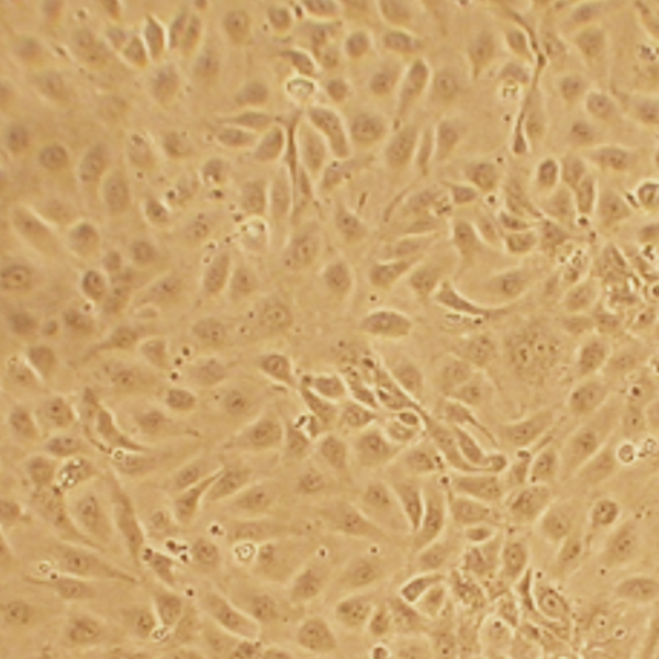
Human Coronary Artery Endothelial Cells: HCAEC-T2D (A). HCAEC stained with DiI-Ac-LDL, the acetylated apoprotein specifically recognized & endocytosed by endothelial cells (B). HCAEC transfected with GFP plasmid DNA using Cytofect™ Endothelial Cell Transfection Kit (C&D).
Human Coronary Artery Endothelial Cells: Type 2 Diabetes: HCAEC-T2D
Human Coronary Artery Endothelial Cells-Plaque (HCAEC-q) are primary macrovascular cells isolated from the human coronary arteries from donors with Type 2 Diabetes.
Description
Human Coronary Artery Endothelial Cells from donors with Type 2 Diabetes (HCAEC-T2D) provide a physiologically grounded model for studying cardiovascular disease at the level of the endothelium — where blood vessel–resident vascular biology is continuously integrated with metabolic cues, mechanical forces from blood flow, and inflammatory signaling. Because endothelial cells are specialized for their vascular bed, coronary artery endothelial cells are particularly relevant for interrogating the mechanisms that distinguish diabetic pathology in the heart from that in other circulations. In this context, HCAEC-T2D capture disease-relevant features associated with type 2 diabetes mellitus and the chronic exposure patterns characteristic of diabetic patients, enabling focused investigation of how diabetes reshapes endothelial dysfunction and promotes coronary atherosclerosis and related cardiovascular events.
In type 2 diabetes, endothelial dysfunction emerges as a central driver linking diabetes to cardiovascular disease, including coronary artery disease, coronary heart disease, and outcomes such as myocardial infarction. High glucose and dysregulated plasma glucose levels contribute to a vascular environment that favors oxidative stress, impaired nitric oxide bioavailability, and heightened endothelial activation. Functionally, this translates into altered vascular inflammation, increased pro-adhesive and pro-chemotactic signaling capacity, and a shift in the balance of endothelial function toward vascular dysfunction. For coronary arteries, these diabetes-associated changes are expected to accelerate coronary atherosclerosis by supporting inflammatory recruitment of circulating immune cells and by reinforcing vascular remodeling programs that can involve cross-talk between endothelial dysfunction and adjacent vascular smooth muscle cells.
HCAEC-T2D are also well suited for examining diabetes-specific signaling mechanisms that underlie coronary angiography–relevant disease phenotypes, including differences between patients with diabetes and non-diabetic patients under comparable experimental conditions. This includes molecular pathways that connect insulin resistance to endothelial activation and altered metabolic response to glucose. Within the coronary microenvironment, insulin resistance and diabetes-associated metabolic stress can amplify endothelial inflammatory tone and reduce the capacity of the endothelium to resolve injury, thereby increasing susceptibility to cardiovascular events. Importantly, studies using coronary artery endothelial cells from diabetic patients have helped clarify how glucose levels and metabolic risk factors converge on endothelial readouts that correlate with coronary atherosclerosis severity and disease progression.
HCAEC-T2D further support mechanistic testing of therapeutic hypotheses relevant to diabetes care and cardiovascular risk reduction, including the impact of metformin and other diabetes interventions on endothelial dysfunction. By probing how these treatments influence vascular inflammation, oxidative stress pathways, and endothelial signaling states, investigators can relate cellular effects to clinical concepts emphasized in medical guidelines and research frameworks such as those promoted by the American Diabetes Association. In this way, HCAEC-T2D connect diabetes mellitus biology to cardiovascular risk factor profiles that include blood pressure and broader cardiometabolic disease context.
Finally, consistent with the broader principle of vascular heterogeneity, endothelial cells from different vascular beds can respond differently to inflammatory cues. Comparing HCAEC-T2D with endothelial cells from other sources helps explain why diabetes-related vascular dysfunction may vary in intensity across vascular territories and why specific stimuli can preferentially promote coronary pathology compared with other vascular disease contexts. Using well-defined primary cells from donors with type 2 diabetes also strengthens interpretability by reducing donor-to-donor confounding such as genetic variability or lifestyle-associated differences, making it more feasible to attribute changes in endothelial function, endothelial dysfunction, and vascular biology readouts to diabetes-driven mechanisms rather than uncontrolled variability.
Human Coronary Artery Endothelial Cells (HCAEC) from Cell Applications, Inc. provide an excellent model system to study all aspects of cardiovascular function and disease, and they have been utilized in dozens of research publications, for example to:
- Understand the mechanism of the anti-inflammatory properties of HDL, and demonstrate for the first time that mature miRNA can control gene expression in a cell where it is neither transcribed nor processed
- Study mechanisms of angiogenesis, as well as oxidative stress and inflammation related pathways in endothelia, including gender and race specific differences in patients with peripheral artery disease
- Elucidate molecular mechanisms of various cardiovascular risk factors, including those associated with diabetes
- Understand the mode of action and cardiovascular protection effects of various natural compounds, vitamins and drug candidates
- Develop and evaluate scaffolds and hydrogels for cardiac tissue engineering, and new treatment strategies to prevent stent restenosis
- Compare effects of BMP-4 on HCAEC and Human Pulmonary Artery Endothelial Cells (HPAEC, also from Cell Applications, Inc.)
- Show that only in HCAEC BMP-4 treatment induced ROS, activated NF-kB, ICAM-1 and increased monocyte adhesiveness, explaining why its upregulation leads to atherosclerosis and hypertension in the systemic, but not pulmonary circulation
Additionally, HCAEC, along with human aortic (HAOEC), carotid artery (HCtAEC), subclavian artery (HScAEC) and brachiocephalic artery (HBcAEC), all provided by Cell Applications, Inc., have been used to demonstrate that not only blood vessels from different tissues are highly heterogeneous, they also interact differently with leukocytes during the inflammation response. The authors further showed that differential N-glycosylation of commonly expressed vascular adhesion molecules may be responsible for this heterogeneity, as well as for modulation of signaling under resting and activated inflammatory conditions. This also explains why specific vascular beds may be more or less susceptible to particular diseases or stimuli. Importantly, if cells from different sources were used, these results could not be convincingly validated due to a number of uncontrolled variables, such as age, race, genetic variability or life style choices of the donors. To eliminate the donor-to-donor variability, the scientists took advantage of the great variety of primary cells offered by Cell Applications, including the option of ordering a panel of endothelial cells obtained from different vascular beds of the same donor!
Because of the complex heterogeneity that exists not only between different donors, but even between different vascular beds in the same individual, it would be prudent to confirm any new findings on primary cell lots coming from several different origins.
Details
| Tissue | Human coronary artery from donor with Type 2 Diabetes (T2D) | |
|---|---|---|
| QC | No bacteria, yeast, fungi, mycoplasma, virus | |
| Character | Factor VIII-related Ag, DiI-Ac-LDL uptake | |
| Bioassay | Attach, spread, proliferate in Growth Med | |
| Cryovial | 500,000 HCAEC-T2D (2nd passage) frozen in Basal Medium w/ 10% FBS, 10% DMSO | |
| Kit | Cryovial frozen HCAEC-T2D (300T2D-05a), Growth Med (212-500), Subculture Rgnt Kit (090K) | |
| Proliferating | Shipped in Gr Med, 3rd psg (flasks or plates) | |
| Doublings | At least 15 | |
| Applications | Laboratory research use only (RUO). Not for human, clinical, diagnostic or veterinary use. |
Resources
FAQs
Need More Help?
Visit our comprehensive FAQ page for detailed answers to common questions.
Need More Help?
Visit our comprehensive FAQ page for detailed answers to common questions.
Primary Cell FAQs Powered by Bioz