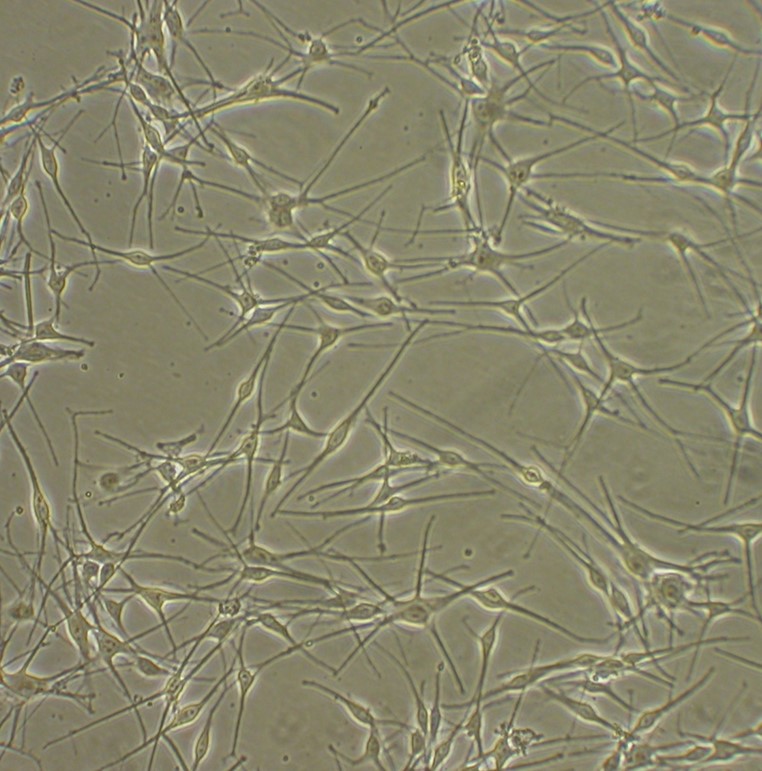
Human Epidermal Melanocytes: HEM. Pigment producting cells isolated from skin tissue.
Human Epidermal Melanocytes: HEM
Human Epidermal Melanocytes (HEM) are highly specialized, neuroectoderm-derived dendritic cells responsible for the synthesis of the protective pigment known as melanin.
Description
Human Epidermal Melanocytes (HEM) are highly specialized, neuroectoderm-derived dendritic cells responsible for the synthesis of the protective pigment known as melanin. Within intact human skin, these pigment-producing cells reside at the basal layer of the epidermis, interspersed among basal keratinocytes. Each individual melanocyte extends elongated cytoplasmic projections, termed dendrites, to physically interact with surrounding epidermal cells. In human skin, the commonly cited ratio is approximately 30 to 40 keratinocytes per single melanocyte, though this exact proportion varies significantly by anatomical site and individual phototype. This highly coordinated anatomical structure is operationally defined as the epidermal melanin unit.
Through this structural unit, synthesized melanin is securely packaged into specialized, lysosome-related organelles called melanosomes. These vesicles are actively transported along the dendritic network and transferred into the cytoplasm of the adjacent keratinocyte population. Once inside, the pigment aligns supranuclearly around the nucleus to provide protection from mutagenic ultraviolet (UV) radiation. This vital process governs baseline and adaptive skin pigmentation.
Beyond interfollicular skin, melanocytes also locate to the bulge region and bulb of the hair follicle, driven by tightly regulated neural crest developmental pathways. To secure an authentic, non-transformed biological baseline for research, primary HEM are typically isolated from human skin tissue biopsies and maintained in highly specialized, serum-free melanocyte growth medium. When maintained in optimized melanocyte growth medium, primary cultured melanocytes maintain their characteristic dendritic shape for multiple generations, offering a useful cell model for tracking proliferation, differentiation, and the earliest signaling alterations driving oncogenic transformation.
While primary cultures bypass the genetic mutations present in immortalized or malignant lines, investigators must note that their population doublings are finite proliferative. Over extended passaging, cultured melanocytes can exhibit progressive culture-induced senescence, a gradual retraction of dendrites, and an altered baseline melanin output. Furthermore, because primary cell viability and biochemical responses are influenced by donor-specific clinical attributes — including age, phototype (Fitzpatrick skin type), and anatomical harvest site—experimental endpoints must be cross-validated across multiple donor lots.
To satisfy international biological safety standards for human-derived tissue, primary HEM lots must be thoroughly screened and verified negative for major blood-borne pathogens, including human immunodeficiency virus (HIV), hepatitis b virus, and hepatitis c virus. Interestingly, beyond conventional dermatological workflows, primary HEM serve as a valuable translational cell model for muscular disease. Because normal human melanocytes endogenously express the full-length muscular isoform of dystrophin at their plasma membrane, specialized studies utilize patient-derived HEM as an accessible, minimally invasive alternative to traditional muscle biopsies to evaluate real-time membrane alterations and downstream mitochondrial dysfunction associated with duchenne muscular dystrophy.
Primary human skin melanocytes provide an essential experimental control and mechanistic model system in peer-reviewed literature, particularly across molecular oncology and pigmentary disorder workflows designed to map cellular senescence, melanocyte migration, and malignant neoplasia progression:
- BRAF Mutations and Melanoma Oncogenesis: A primary application of HEM is defining the central role of oncogenic driver genes in skin cancer development. Investigators use HEM to study how a single somatic mutation in the BRAF gene (most notably the B-RAFV600E hotspot) robustly activates the mitogen-activated protein kinase (MAPK) pathway. Literature reports indicate that this constitutive activation can drive downstream interactions with the mechanistic target of rapamycin (mTOR) pathway, STAT3-dependent transcription of the anti-apoptotic protein Mcl-1, and Tbx3-mediated repression of E-cadherin. However, researchers must note that these downstream impacts involve context-specific and often indirect links rather than inevitable, single-step outcomes. When these complex pathways align, they are reported to alter cell adhesion, accelerate melanocyte migration, and contribute to heightened metastatic potential, making HEM a foundational benchmark to identify unique features and early biomarkers of melanoma cells.
- Mechanisms of Cellular Senescence and Tumor Suppression: HEM models are heavily utilized to dissect the molecular checkpoints that govern cellular aging and suppress tumorigenesis. Investigators leverage cultured melanocytes to show that overexpressing the inhibitor of DNA binding 1 (Id1) protein can extend the replicative lifespan of melanocytes through the regulation of p16INK4a tumor suppressor expression, though these effects remain highly cell- and model-dependent. In parallel oncogenic workflows, HEM have been deployed to demonstrate that the p16INK4a-Rb-CDK4/6 senescence-inducing pathway is frequently inhibited or bypassed in melanoma cells, permitting the unrestrained proliferation of cells harboring severe DNA damage. Conversely, specific senescence modeling in HEM has shown that insulin-like growth factor-binding protein 7 (IGFBP7) is context-dependent and not universally required for B-RAFV600E-induced senescence across all experimental backgrounds.
- Complementary Oncogenic Interplay and Metabolic Shifts: Beyond the canonical MAPK cascade, primary HEM are essential for identifying cooperating signaling partners in melanoma development. Researchers use these cells to track how phosphoinositide 3-kinase (PI3K) participates in regulating MAPK activation in response to oncogenic c-Kit receptor tyrosine kinase activity, and how the Nck2 adaptor protein coordinates intracellular tyrosine kinase networking. Furthermore, comparative metabolic profiling between normal HEM and advanced melanoma lines has revealed a definitive metabolic shift. Melanoma progression often involves increased glycolysis (the Warburg effect) and altered mitochondrial function, though the exact extent and timing of this metabolic reprogramming vary considerably by tumor stage and histological subtype.
- Melanocyte Migration and Stemness Dynamics: Regulating the movement of melanocytes is critical during both embryonic development and post-inflammatory hyperpigmentation. Researchers utilize primary HEM inside specialized migration chambers to evaluate how localized growth factor gradients, extracellular matrix components, and ultraviolet stress accelerate or blunt real-time melanocyte migration. Furthermore, in the context of tissue regeneration and skin pigmentation restoration, HEM serve as a human comparative model alongside undifferentiated neural crest stem cell populations, allowing investigators to map the precise transcription factors and environmental cues that compel an uncommitted stem cell to differentiate into a mature, melanin-producing epidermal melanocyte.
Human Epidermal Melanocytes (HEM) from Cell Applications, Inc. maintain their characteristic shape in culture for many generations. They produce melanin and serve as a useful cell model for the studies of melanocyte proliferation and differentiation, as well as progression of melanocytic neoplasia. Epidermal melanocytes are pigment-producing cells located at the basal level of epidermis, where they interact with keratinocytes via cellular processes called dendrites. Melanin, the pigment produced by melanocytes and responsible for skin color, is then transferred to keratinocytes, where it is stored in vesicles called melanosomes located around the nucleus to provide protection from UV radiation.
HEM provided by Cell Applications, Inc. have been used to:
- Identify unique features and biomarkers of melanoma cells
- Show that IGFBP7 is dispensable for B-RAFV600E-induced senescence
- Investigate mechanisms of cellular senescence, in particular by showing that Id1 extends the life span of melanocytes through inhibition of p16INK4a expression
- Discover the central role of oncogenic BRAF gene in melanoma oncogenesis by demonstrating that the constitutive MAPK pathway activation leading to activation of mTOR, STAT3-dependent transcription of Mcl-1, Tbx3-mediated repression of E-cadherin leading to increased metastasis in melanoma cells all result from overexpression and/or mutations of BRAF gene
- Discover other key players in oncogenic signaling leading to melanoma, such as PI3K which regulates MAPK activation in response to oncogenic c-Kit activity, Nck2 adaptor protein which participates in regulation of tyrosine kinases activity and the role of mitochondria metabolism in advanced melanoma
- Demonstrate that p16INK4a-Rb-CDK4/6 senescence-inducing tumor suppressor pathway in inhibited in melanoma cells leading to proliferation of cells harboring DNA damage and by studying expression of senescence markers
Details
| Tissue | Normal healthy human neonatal foreskin | |
|---|---|---|
| QC | No bacteria, yeast, fungi, mycoplasma, virus | |
| Bioassay | Attach, spread, proliferate in Growth Med | |
| Cryovial | 500,000 HEM (2nd passage) frozen in Basal Medium w/ 30% FBS, 10% DMSO | |
| Kit | Cryovial frozen HEM (104-05n), Growth Medium (135-500), Subcltr Rgnt Kit (090K) | |
| Proliferating | Shipped in Gr Med, 3rd psg (flasks or plates) | |
| Doublings | At least 12 | |
| Applications | Laboratory research use only (RUO). Not for human, clinical, diagnostic or veterinary use. |
Resources
FAQs
Need More Help?
Visit our comprehensive FAQ page for detailed answers to common questions.
Need More Help?
Visit our comprehensive FAQ page for detailed answers to common questions.
Primary Cell FAQs Powered by Bioz