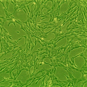
Canine Chondrocytes: CnC
Canine Chondrocytes (CnC) are highly differentiated, specialized cells isolated from the healthy articular cartilage of canine (Canis lupus familiaris) joints.
Description
Canine Chondrocytes (CnC) — alternatively designated as a primary canine chondrocyte or articular chondrocyte population — are highly differentiated, specialized cells isolated from the healthy articular cartilage of canine (Canis lupus familiaris) joints. In mature articular cartilage, these cells function as the sole resident cell type, responsible for the ongoing synthesis, structural organization, and metabolic maintenance of a dense, extracellular matrix (ECM) rich in type II collagen and abundant sulfated proteoglycans like aggrecan. Structurally, each cell natively resides within lacunae surrounded by a specialized pericellular matrix in vivo. Their population-doubling capacity is finite, but unlike a transformed cell line or immortalized cell isolates, primary CnC retain their native signaling networks and diploid integrity when expanded in standard culture media.
In the living organism, this specific cell type is distributed sparsely throughout the unmineralized and mineralized layers of articular cartilage lining the load-bearing surfaces of diarthrodial joints. Because healthy mature cartilage is largely avascular and poorly innervated, these cells rely entirely on passive nutrient diffusion through the surrounding matrix from the synovial fluid, while sensory and vasoactive nerve fibers remain restricted to the surrounding joint capsule and subchondral bone. While adult stem cells — such as multipotent canine mesenchymal stem cells (canine MSCs) or mesenchymal stromal cells isolated from adipose tissue or bone marrow — can undergo chondrogenic differentiation to generate cartilage-like cells, primary CnC are harvested directly from fully committed joint tissues. Given the remarkably high clinical prevalence of degenerative joint disease and osteoarthritis in canine populations — particularly within large breeds and geriatric dogs — sourcing primary cells from healthy donor joints provides a translationally invaluable comparative model for both veterinary and human orthopedic medicine, frequently contrasted with human MSCs in cross-species studies.
The primary biological function of a canine chondrocyte is to govern cartilage homeostasis by maintaining a meticulous balance between matrix synthesis (anabolism) and matrix degradation (catabolism). Under physiological loading, they secrete structural proteins and glycosaminoglycans to withstand mechanical compression and fluid shear inside the joint. However, their experimental utility is profoundly shaped by the dimensionality of their culture environment. When expanded in standard two-dimensional (2D) monolayer cultures on plastic, CnC exhibit a high propensity for culture-induced “dedifferentiation.” This drift is characterized by a morphological shift from a rounded, native chondrocytic shape to an elongated, fibroblast-like appearance, which downregulates the master transcription factor SOX9 and cartilage-specific matrix proteins, while upregulating type I collagen and fibronectin expression. To circumvent this, researchers utilize canine chondrocyte basal medium paired with 3D encapsulation systems like alginate microspheres or biomimetic hydrogels to sustain a rounded morphology and support appropriate type II collagen deposition.
In veterinary pharmacology, molecular biology, and tissue engineering, CnC represent a premier screening platform to validate the safety, antioxidant properties, and mechanisms of anti-arthritic compounds. Investigators deploy these cells to evaluate the cytoprotective activities of commercial supplements, tracking shifts in gene expression via qPCR and monitoring how effectively they blunt inflammatory surges canonically induced via interleukin-1β (IL-1β) challenge. In these models, researchers measure the downregulation of destructive matrix metalloproteinases (MMPs) and cyclooxygenase-2 (COX-2) activity using protein expression analyses like Western blot or ELISA, while evaluating protective effects through functional assays like biochemical glycosaminoglycan (GAG) quantification and mechanical testing of the engineered tissue.
Furthermore, due to the highly restricted innate capacity of cartilage for self-repair, bioengineers widely pair CnC with novel biomaterial scaffolds, micropatterned surfaces, and microfluidic lab-on-a-chip devices to develop engineered tissue solutions and evaluate cartilage repair strategies for focal cartilage defects. While high-throughput veterinary therapeutic discovery pipelines often utilize simpler automated readouts such as cell viability or reporter gene assays to screen large compound libraries, secondary characterization relies on a multi-faceted approach. Investigators routinely combine flow cytometry (to verify cell surface marker profiles) with histology, biochemical assays, and gene expression profiling to comprehensively assess cell phenotype stability and matrix production before moving toward cell transplant modeling.
Details
| Tissue | Normal healthy canine articular cartilage | |
|---|---|---|
| QC | No bacteria, yeast, fungi, mycoplasma | |
| Bioassay | Attach, spread, proliferate in Growth Med | |
| Cryovial | 500,000 CnC (1st passage) frozen in Basal Medium w/ 10% FBS, 10% DMSO | |
| Kit | Cryovial frozen CnC (Cn402-05), Growth Medium (Cn411-500), Subcltr Rgnt Kit (090K) | |
| Proliferating | Shipped in Gr Med, 2nd psg (flasks or plates) | |
| Doublings | At least 10 | |
| Applications | Laboratory research use only (RUO). Not for human, clinical, diagnostic or veterinary use. |
Resources
FAQs
Need More Help?
Visit our comprehensive FAQ page for detailed answers to common questions.
Need More Help?
Visit our comprehensive FAQ page for detailed answers to common questions.
Primary Cell FAQs Powered by Bioz