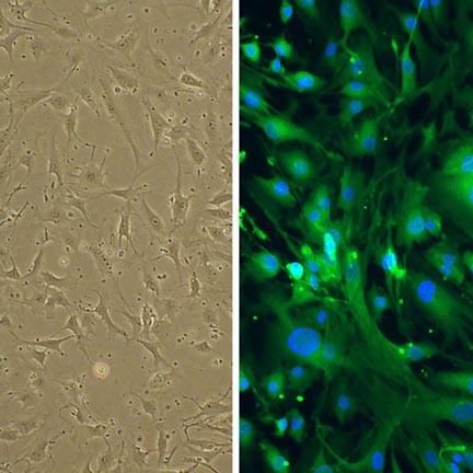
Human Astrocytes: HA
Human Astrocytes (HA) are specialized glial cells (or astroglia) that constitute one of the most abundant glial cell type in the central nervous system (CNS).
Description
Human Astrocytes (HA) are isolated from human cerebral tissue. Astrocytes play vital homeostatic roles essential for neurovascular coupling, structural architecture, metabolic support, and synaptic orchestration. HA are specialized glial cells (or astroglia) that constitute one of the most abundant glial cell type in the central nervous system (CNS). As a true primary cell type rather than modified immortalized cell lines, these human primary cells preserve native genetic backgrounds and realistic physiological responsiveness. Characterization of a primary astrocyte culture verifies that they maintain typical astrocyte surface molecule expression, distinct starburst morphologies, and localized signaling networks that are frequently lost or altered in continuous cultures. To maximize cumulative population doublings and minimize unguided activation or fibroblastic overgrowth in vitro, investigators rely on a highly optimized, serum-free astrocyte medium, though serum-free formulations reduce rather than fully prevent reactive changes.
In the mammalian brain and spinal cord, these vital glial cells are ubiquitously distributed throughout both grey and white matter, positioning themselves structurally between neuronal networks and the cerebral microvasculature. While commercial cell systems provide discrete, region-specific lots — such as human astrocytes hippocampal, human astrocytes cerebellar, or cells sourced from the human midbrain (mesencephalon) — astrocyte heterogeneity is highly complex. Functional diversity is shaped not only by gross anatomical labels, but also by overlapping molecular subtypes, developmental stages, and localized reactive states. Across all regions, the astrocyte cell extends specialized cytoplasmic processes, known as end-feet, that form extensive endfoot contacts with cerebral vessels. These end-feet extensively ensheath brain capillaries, working alongside other integral components — such as endothelial cells, pericytes, and the vascular basement membrane — to form a fully integrated neurovascular unit.
The primary biological function of healthy astrocytes is to maintain CNS homeostasis, provide structural architecture, and manage neurovascular coupling. Energetically, they provide metabolic support to neurons; mechanisms such as the astrocyte-neuron lactate shuttle represent a widely supported model for metabolic provision, though it remains debated in certain physiological contexts. At the neurovascular interface, they provide reciprocal signaling to help establish and maintain the blood brain barrier. While the physical restriction of immune cell trafficking is primarily a property of the endothelial barrier and its tight junctions, astrocytes contribute essential modulatory signals that regulate the recruitment and signaling of peripheral immune entities like a circulating T cell or dendritic cell. Furthermore, they are active participants in synaptic plasticity, clearing excess neurotransmitters (such as glutamate) from the synaptic cleft to minimize excitotoxicity, and cooperating with microglia to manage synaptic pruning. During development or following injury, they also secrete neurotrophic factors that influence neurite outgrowth, provide neuronal guidance, and support neuron survival and neuron regeneration.
In laboratory settings, Human Astrocytes (HA) provide an indispensable human-background platform for exploring functional astrocyte diversity, tracking pathological processes, and evaluating neuroprotective pharmacology. Numerous studies utilize HA alongside glioblastoma cell lines to identify unique transcriptomic signatures, discover target biomarkers, and optimize diagnostic positron emission tomography (PET) imaging for high-grade gliomas. In neurodegenerative disease modeling, researchers expose these cells to pathological protein aggregates — such as -synuclein pre-formed fibrils — and pro-inflammatory cytokine cocktails to map out shifts in gene expression and visualize mitochondrial bioenergetic collapse via high-resolution immunofluorescence. While these cells are intended strictly for in vitro research use, they serve as a rigorous testing ground for nanomedicine development, allowing bioengineers to evaluate the target specificity and safety of core-cross-linked nanoparticles designed to mitigate neuroinflammation or cross disrupted physiological barriers.
- Investigate aberrant gene expression.
- Reveal that CBX6 overexpression inhibits proliferation of glyoblastoma cells and
- Demonstrate the role of epigenetics in glyioblastoma malignancy.
- Investigate choline uptake system in normal and glioblastoma cells with the hope to improve tomography imaging in glioblastoma patients
Details
| Tissue | Normal healthy Human brain | |
|---|---|---|
| QC | No bacteria, yeast, fungi, mycoplasma, virus | |
| Character | Positive for GFAP | |
| Bioassay | Attach, spread, proliferate in Growth Medium | |
| Cryovial | 500,000 HA (2nd passage) frozen in Basal Medium w/10% FBS, 10% DMSO | |
| Kit | Cryovial frozen HA (882A-05), Growth Medium (821-500), Subculture Rgnt Kit (090K) | |
| Proliferating | Shipped in Gr Med, 3rd psg (flasks or plates) | |
| Doublings | At least 10 for fetal, at least 8 for adult. | |
| Applications | Laboratory research use only (RUO). Not for human, clinical, diagnostic or veterinary use. |
Resources
FAQs
Need More Help?
Visit our comprehensive FAQ page for detailed answers to common questions.
Need More Help?
Visit our comprehensive FAQ page for detailed answers to common questions.
Primary Cell FAQs Powered by Bioz