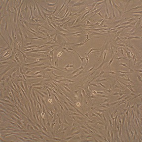
Human Fibroblast-Like Synoviocytes: HFLS
Human Fibroblast-Like Synoviocytes (HFLS) are specialized mesenchymal stromal cells isolated from the synovial membrane (synovium) lining joint cavities.
Description
Human Fibroblast-Like Synoviocytes (HFLS) — also frequently designated as synovial fibroblasts — are specialized mesenchymal stromal cells isolated from the synovial membrane (synovium) lining joint cavities. Structurally characterized by a spindle-shaped morphology, these fibroblast-like synoviocytes are primary architectural elements of the synovial tissue. Unlike continuous, transformed cell lines, primary HFLS maintain an anatomically primed memory of their tissue of origin, providing a more translationally rigorous baseline. To preserve their specialized phenotype and support post-thaw recovery in vitro, they are typically cultured in optimized environments, utilizing optimized HFLS Growth Medium or a specialized HFLS basal medium with a defined HFLS growth supplement according to the protocol.
Beyond their classical use in joint biology, primary HFLS from Cell Applications hold profound historical significance in the fields of regenerative medicine and developmental biology. Notably, along with Human Dermal Fibroblasts (HDF) from Cell Applications, primary Human Fibroblast-Like Synoviocytes (HFLS) from Cell Applications served as a critical human somatic donor cell source in the seminal, landmark research published in Cell and related journals in 2007 by Dr. Shinya Yamanaka and colleagues. This breakthrough work demonstrated that the retroviral transduction of four core transcription factors (, , , and ) could successfully reprogram somatic cell types back into a pluripotent state — a discovery that established the foundation of induced pluripotent stem cell (iPSC) technology and for which Dr. Yamanaka received the Nobel Prize in Physiology or Medicine in 2012.
In the living organism, these stromal cells are localized within the synovium, a specialized soft connective tissue that lines the non-cartilaginous surfaces of diarthrodial joints. High-resolution single-cell mapping studies highlight that the synovial membrane contains distinct anatomical fibroblast subsets. These are broadly categorized into those residing in the intimal lining layer — which directly interfaces with the synovial fluid and expresses specific surface markers like podoplanin and cadherin-11 — and those located in the deeper sublining layer, which can be characterized by a distinct phenotype.
While macrovascular or dermal fibroblasts manage their respective localized barriers, joint-derived HFLS are anatomically and microenvironmentally adapted to withstand mechanical articulation forces and govern the strict biochemical constraints of the joint space.
The primary biological function of healthy HFLS is to maintain joint homeostasis, provide structural support, and regulate nutrient exchange within the articular cavity. In a healthy physiological state, these cells synthesize the underlying extracellular matrix and produce key lubricating macromolecular elements of the synovial fluid, such as hyaluronic acid and lubricin, and are involved in regulating the localized immune environment.
However, during the progression of severe chronic arthropathies—such as rheumatoid arthritis (RA) or osteoarthritis (OA) — their functional baseline undergoes a dramatic pathobiological shift. In response to an altered, inflamed joint environment, HFLS can transition from homeostatic regulators to active contributors to disease progression. They can assume an activated state characterized by a hyperproliferative, apoptosis-resistant phenotype. Though this behavior is a phenotypic analogy and these cells are not cancerous, this automated inflammatory response is associated with an increased interaction with invading immune cells, which can contribute to a self-sustaining loop of localized tissue inflammation and damage.
In cell biology, molecular biology, immunology, and discovery pharmacology, HFLS serve as an indispensable human-background model system for exploring normal joint physiology, uncovering complex RA pathogenesis mechanisms, and evaluating potential anti-rheumatic therapeutics. Historically, pioneering research by investigators like Firestein GS established the critical role these cells play in joint pathology. Researchers routinely stimulate primary HFLS with macrophage-derived pro-inflammatory cytokines (such as TNF- and IL-1β) to study downstream cytokine-driven matrix degradation and map how these signals alter intracellular pathways, cell proliferation rates, and non-coding RNA networks.
In many experimental setups, exposure to an inflammatory cytokine cocktail can induce HFLS to produce inflammatory mediators and matrix-degrading enzymes, such as matrix metalloproteinases (MMPs) and disintegrins (ADAMTS), which are studied in relation to cartilage degradation models. Because these primary cells can retain their disease-primed memory ex vivo, pharmaceutical discovery pipelines deploy them to evaluate novel small-molecule inhibitors, test targeted biologics, and validate therapeutic interventions designed to attenuate inflammatory responses in chronic arthropathies. Finally, due to their established role in somatic cell reprogramming, these cells can be utilized in stem cell research methodologies focused on somatic cell behavior, reprogramming efficiency, and the development of pluripotent lines.
- TNFα – mediated changes in gene expression
- TNF-like weak inducer of apoptosis (TWEAK) contribution to joint inflammation by inducing chemokines and matrix metalloprotease-9, suggesting TWEAK as a new therapeutic target
- The role of miR-124a in arthritis pathogenesis
- C/EBPβ regulation of metalloproteinases and ADAMTS family members in synoviocytes stimulated with IL-1β
- HMW-HA suppression of ADAMTS4 mRNA and protein expression via CD44, p38 MAPK and JNK pathways
- Anti-inflammatory and antirheumatic activity of various compounds, such as NF-κB inhibitors, AGIX-4207, HA–methotrexate conjugates and bucillamine
- Reactive arthritis triggered by chlamydial infection
- Mechanisms of arthritis-like syndrome in patients infected with chikungunya (CHIK) virus
- Use of Leukocyte-poor, RBC-free platelet-rich plasma in clinical orthopaedics, because leukocytes and RBCs cause synoviocyte death and proinflammatory mediator production
- Cartilage link protein and MAGP2 as markers to distinguish chondrocytes and synovial cells
- Creation of induced pluripotent stem cells (iPSC) by using the now classic “Yamanaka cocktail”, the discovery for which Dr. Shinya Yamanaka was awarded the Nobel Prize in 2012
- Causative agents, immunopathological mechanisms and signal transduction pathways leading to joint inflammation in rheumatoid arthritis
- The role of estrogen signaling in increasing inflammation
- Anti-inflammatory properties of herbal compound Sinomenine suggested for rheumatoid arthritis treatment
- Effects of extracellular matrix composition on cell attachment and migration relevant to T-cell function in inflamed tissues
- The role of human endogenous retroviruses (HERVs) in development of rheumatoid arthritis
- Activated expression of different forms of HERV, contributing to development of rheumatoid arthritis symptoms by different mechanisms
Details
| Tissue | Normal healthy human synovial tissue | |
|---|---|---|
| QC | No bacteria, yeast, fungi, mycoplasma, virus | |
| Bioassay | Attach, spread, proliferate in Growth Med | |
| Cryovial | 500,000 HFLS (2nd passage) frozen in Basal Medium w/ 10% FBS, 10% DMSO | |
| Kit | Cryovial frozen HFLS (408-05a), Growth Medium (415-500), Subcltr Rgnt Kit (090K) | |
| Proliferating | Shipped in Gr Med, 3rd psg (flasks or plates) | |
| Doublings | At least 5 | |
| Applications | Laboratory research use only (RUO). Not for human, clinical, diagnostic or veterinary use. |
Resources
FAQs
Need More Help?
Visit our comprehensive FAQ page for detailed answers to common questions.
Need More Help?
Visit our comprehensive FAQ page for detailed answers to common questions.
Primary Cell FAQs Powered by Bioz