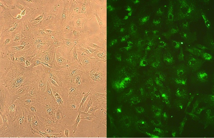
Human Lung Fibroblasts: HLF
Human Lung Fibroblasts (HLF) are primary fibroblast cells isolated from healthy parenchymal lung tissue.
Description
Human Lung Fibroblasts (HLF) are primary cells isolated from healthy parenchymal lung tissue. As resident mesenchymal cells of the respiratory architecture, these primary lung fibroblast isolates are characterized in vitro by an elongated, spindle-shaped morphology and a specialized capacity to synthesize structural matrix proteins. Unlike transformed cell lines or highly mutated cancer cells, normal human lung fibroblasts maintain an intact diploid genome, native receptor baselines, and physiologically authentic signaling cascades. However, because they are primary human fibroblast cell systems, they have a finite lifespan in cell culture and are highly sensitive to their mechanical and biochemical microenvironment. To preserve their native phenotypic properties and avoid premature senescence, they are typically stored long-term as frozen cells and must be maintained in optimized fibroblast growth media. Quality control of these diverse primary cells requires meticulous screening to ensure they are free of mycoplasma and to verify baseline purity compared to other structural cell type contaminants.
In the living organism, this cell type is distributed extensively throughout the lung interstitium and within the delicate alveolar septa that separate adjacent air spaces. Within this specific alveolar-capillary microenvironment, pulmonary fibroblasts reside in close anatomical and paracrine proximity to alveolar epithelial cells (Types I and II), capillary endothelial cells, and resident immune cells. While multipotent stem cell populations or circulating mesenchymal progenitors can home to the respiratory tract during severe injury, resident HLF remain the principal cell type responsible for maintaining daily alveolar wall architecture, regulating local tissue compliance, and coordinating structural homeostatic maintenance.
The primary biological function of a lung fibroblast is to govern the composition and turnover of the pulmonary extracellular matrix (ECM) by meticulously balancing the synthesis of structural elements —such as types I, III, and V collagen, fibronectin, and elastin — with the secretion of matrix metalloproteinases (particularly MMP-1, MMP-2, and MMP-9) and tissue inhibitors of metalloproteinases (TIMPs). However, in response to direct tissue injury, chronic inflammation, or pathological cues, these quiescent/homeostatic fibroblasts undergo a profound functional transition known as myofibroblast differentiation.
Triggered by mechanical tissue rigidity or local growth factors like transforming growth factor-beta 1 (TGF-1), this shift drives the reorganization of the actin cytoskeleton, assembly of -SMA–positive stress fibers, and heightened collagen contraction. While local proliferation of resident homeostatic fibroblasts is a primary driver of this contractile pool, other sources — including resident pericytes, partial epithelial-to-mesenchymal transition (EMT)/transdifferentiation, and bone-marrow–derived fibrocytes — can also contribute to the myofibroblast population in vivo, though the extent of full EMT contribution remains a subject of ongoing debate. While temporary myofibroblast activity is required for effective tissue repair and normal wound healing, its persistence under chronic pathological stimulation results in abnormal tissue function. This drives chronic ECM deposition — where collagens I and III serve as the dominant fibrillar collagens in fibrosis, while collagen V is present but less abundant — and persistent architectural remodeling of alveolar septa, which can compromise gas exchange and contribute to secondary complications like pulmonary hypertension via concurrent vascular remodeling.
In pulmonology, molecular biology, and safety pharmacology, HLF cultures function as a premier human-background model system to dissect the pathways driving both physiological lung function and complex respiratory disease states. Investigators deploy these primary cells to map the molecular mechanics of idiopathic pulmonary fibrosis (IPF) and progressive interstitial lung disease, utilizing single-cell transcriptomic (scRNA-seq) and proteomic datasets to identify distinct fibroblast subpopulations within the diseased IPF lung. These single-cell analyses, coupled with emerging spatial transcriptomic studies, highlight significant contextual and spatial heterogeneity among distinct fibroblast lineages. Through these screens, researchers track differential gene expression profiles, mapping how targeted receptor-interacting proteins and altered cytokine or interleukin networks alter cellular expansion and matrix deposition.
Furthermore, HLF serve as an essential translational platform to study oncogenic cross-talk and the tumor microenvironment in lung cancer. In co-culture models, investigators study how native fibroblasts transition into a cancer-associated fibroblast (CAF) phenotype, tracing how tumor-derived factors drive excess proliferation and metabolic reprogramming that supports cancer cell survival. Beyond oncology and fibrotic disease, primary HLF are heavily utilized in discovery pharmacology and toxicological screening. They are essential for modeling cell behavior changes under hypoxic injury, testing novel antifibrotic or anti-angiogenic small molecules, and evaluating targeted therapies engineered to achieve effective tissue repair and halt pathological tissue expansion without inducing unwanted cellular cytotoxicity.
- Differential gene expression, cytokines, growth factors and interleukins
- Receptor activation, inhibition and receptor-interacting protein
- Cell behavior changes, including synthesis of collagen and other extracellular matrix molecules, as well as actin reorganization, collagen contraction and fibroblast / myofibroblast trans-differentiation.
- Lung disease, disorders and injury, including hypoxia, tissue rigidity, pulmonary fibrosis and hypertension
- That unwanted cell growth can be inhibited, and that the healing process can be understood and improved
Details
| Tissue | Normal healthy human lung parenchyma | |
|---|---|---|
| QC | No bacteria, yeast, fungi, mycoplasma, virus | |
| Bioassay | Attach, spread, proliferate in Growth Med | |
| Cryovial | 500,000 HLF (1st passage) frozen in in Basal Medium w/ 10% FBS, 10% DMSO | |
| Kit | Cryovial frozen HLF (506-05a or 506-05f), Gr Med (516-500), Subcltr Rgnt Kit (090K) | |
| Proliferating | Shipped in Gr Med, 2nd psg (flasks or plates) | |
| Doublings | At least 12 | |
| Applications | Laboratory research use only (RUO). Not for human, clinical, diagnostic or veterinary use. |
Resources
FAQs
Need More Help?
Visit our comprehensive FAQ page for detailed answers to common questions.
Need More Help?
Visit our comprehensive FAQ page for detailed answers to common questions.
Primary Cell FAQs Powered by Bioz