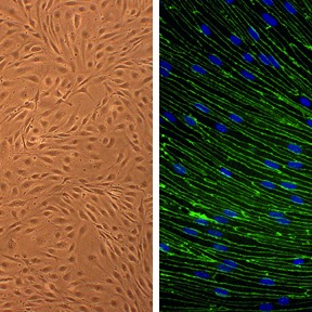
Rat Brain Microvascular Endothelial Cells: RBMVEC
Rat Brain Microvascular Endothelial Cells (RBMVEC) are highly specialized vascular endothelial cells isolated from the continuous capillaries of the rat (Rattus norvegicus) central nervous system (CNS).
Description
Rat Brain Microvascular Endothelial Cells (RBMVEC) — alternatively written as RBMVECs or designated as a primary brain capillary endothelial cell type in the literature — are highly specialized vascular endothelial cells isolated from the continuous capillaries of the rat (Rattus norvegicus) central nervous system (CNS). Unlike a transformed continuous cell line, immortalized epithelial cells, or generic tumor endothelial cell culture groups, these rat primary cells better retain their native in vivo cell biologics, architectural complexity, and strict barrier characteristics ex vivo. To preserve their specific functional properties, prevent dedifferentiation, and maintain high cell viability, investigators rely on specialized Rat Brain Endothelial Cell culture media supplemented with precise concentrations of endothelial cell growth supplements. Quality control validation of these cell biologics products typically includes flow cytometry or immunocytochemical analysis to verify the continuous, peripheral localization of canonical markers such as CD31 (PECAM-1) and VE-cadherin.
In the living organism, these cells form the continuous, non-fenestrated luminal inner lining of the microvessels and capillaries throughout the brain parenchyma. The process of isolating RBMVECs from rat tissues requires microdissecting brain microvessels away from surrounding neural matrices, followed by sequential enzymatic digestion and cell isolation steps. In comparative vascular molecular biology, RBMVEC serve as a foundational rodent model for the blood–brain barrier (BBB). While researchers frequently utilize human primary cells, monkey primary cells, or canine primary cells for species-specific translational benchmarking, these rat-derived brain endothelial cells remain highly valued in neurobiology due to the wealth of matching physiological data available across diverse peer-reviewed journals, such as Brain Res and other specialized neuroscience publications.
The primary biological function of this specialized capillary endothelial cell population is to form the physical and metabolic backbone of the blood–brain barrier, strictly regulating CNS homeostasis and protecting the neural parenchyma from circulating neurotoxins, peripheral immune cells, and fluctuating systemic concentrations. This restrictive barrier capacity is structurally defined by the dense assembly of specialized tight junction complexes — most notably claudins (such as claudin-5), occludin, and zonula occludens-1 (ZO-1) — which physically seal the paracellular space. Additionally, these cells express a distinct repertoire of polarized active efflux transporters belonging to the ATP-binding cassette (ABC) superfamily, including P-glycoprotein (P-gp) and Breast Cancer Resistance Protein (BCRP), which actively extrude a wide array of small molecules back into the vascular lumen.
In preclinical drug discovery, neurotoxicology, and stroke research, RBMVEC cell culture platforms serve as a vital gatekeeping assay to evaluate the brain penetration of novel neuroprotective agents and map pathological permeability cascades. Investigators routinely deploy these cells to model oxygen-glucose deprivation (OGD), assess drug-induced barrier disruption, and test advanced drug delivery vehicles (such as surface-modified nanoparticles) designed to safely bypass the BBB. However, researchers must note that species differences between rats and humans in transporter expression, tight-junction regulation, and downstream signaling pathways can limit direct translational inference. Furthermore, these cells display high culture sensitivity; tight junctions, active transporter expression, and overall barrier function decline rapidly in standard 2D static culture unless actively supported by fluidic shear stress, multi-cellular co-culture, or highly optimized media formulations. To improve the in vivo-like phenotype, protocols increasingly incorporate these physiological parameters alongside primary astrocytes and pericytes to recreate the multi-cellular neurovascular unit (NVU).
To expand experimental capabilities, cell culture services provide specialized modifications, such as custom cell isolation protocols, frozen cells for high-throughput screening, and engineered variants including GFP expressing cells or monolayers tailored for endothelial specific Adenoviral overexpression studies. This flexibility allows researchers to compare outcomes across species by matching RBMVEC data against mouse primary cells, broader mouse tissues, or rabbit cells to ensure a comprehensive overview of mammalian vascular dynamics.
- Serve as a gold standard control for endothelial markers VE-cadherin and CD31 expression
- Demonstrate a better brain penetration of cardiovascular and anti-stroke drugs Tanshinones IIA and IIB and Cryptotanshinone in the presence of PgP or MRP1/2 inhibitors or by using PEGylated gold nanoparticles
- Show that removal of excess glutamate from the blood by glutamate oxaloacetate transaminase correlates to a decrease in brain glutamate levels and confers neuroprotection against ischemic stroke
- Assess, along with Rat Astrocytes (RA) also obtained from Cell Applications, Inc., neuroprotective capabilities of bioenergy stabilizers in an in vitro model of stroke
- Show that bile acids cause activation of Rac1 and phosphorylation of occluding, explaining increased permeability of the blood brain barrier seen during obstructive cholestasis
- Demonstrate that saquinavir-associated dementia in HIV-positive patients is exacerbated by smoking due to the BBB disruptive effects of both nicotine and saquinavir, mediated by decrease in Notch-4 expression and increase in ROS
- Show that treatment with connexin43 mimetic peptide which transiently blocks gap junction function, reduces vascular leak and can be used to treat of central nervous system ischaemia
- Reveal that oxygen and glucose deprivation increase ROS, cytochrome c levels and caspase-3 activity, induce tight junction disruption and actin stress fiber formation, leading to BBB break down
- Investigate the interactions between neurons, astorcytes and endothelial cells by showing that exposure to metabolic stress induces tissue-type plasminogen activator release from neurons which induces AMPK activation, membrane recruitment of GLUT1, and GLUT1-mediated glucose uptake in astrocytes and endothelial cells, followed by the synthesis and release of lactic acid from astrocytes, and that the uptake of this lactic acid via the monocarboxylate transporter-2 promotes survival in neurons
- Study the proapoptotic and anti-angiogenic role of p75NTR in choroidal neovascularization
Details
| Tissue | Normal healthy brain from adult rat | |
|---|---|---|
| QC | No bacteria, yeast, fungi, mycoplasma | |
| Character | DiI-Ac-LDL uptake: Positive | |
| Bioassay | Attach, spread, proliferate in Growth Med | |
| Cryovial | 500,000 RBMVEC (2nd psg) frozen in Basal Medium w/ 10% FBS, 10% DMSO. | |
| Kit | Cryovial RBMVEC (R840-05a), Gr Med (R819-500), Attch Fctr Sln (123-100), Subcltr Rgnt Kit (090K) | |
| Proliferating | Shipped in Gr Med, 3rd psg (flasks or plates) | |
| Doublings | At least 10 | |
| Applications | Laboratory research use only (RUO). Not for human, clinical, diagnostic or veterinary use. |
Resources
FAQs
Need More Help?
Visit our comprehensive FAQ page for detailed answers to common questions.
Need More Help?
Visit our comprehensive FAQ page for detailed answers to common questions.
Primary Cell FAQs Powered by Bioz