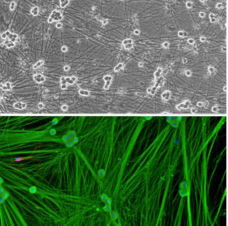
Rat Dorsal Root Ganglion Neurons: RDRGN
Rat Dorsal Root Ganglion Neurons (RDRGN) are primary sensory neurons isolated from the spinal ganglion (dorsal root ganglia) of rats (Rattus norvegicus).
Description
Rat Dorsal Root Ganglion Neurons (RDRGN) — also called rat DRG neurons or studied within broader DRG cultures — are primary sensory neurons isolated from the spinal ganglion (dorsal root ganglia) of rats (Rattus norvegicus). Morphologically, these cells develop as pseudounipolar cells, featuring a single axon that bifurcates into a peripheral branch that innervates target tissues and a central branch that enters the spinal cord. Unlike an immortalized neuronal cell line, these dissociated neurons better preserve their native architectural complexity, receptor repertoires, and electrophysiological properties in a primary culture, though properties can drift with dissociation and time in culture. To maintain their specialized, non-mitotic phenotype and support long-term cell survival in vitro, these sensory nerve cell bodies require highly optimized conditions that differ significantly by neuron subtype and donor age (neonatal vs. adult DRG). Investigators generally cultivate them using neurobasal media supplemented with B27 or NS21, alongside tailored neurotrophin combinations rather than just isolated NGF.
In the living organism, these dorsal root ganglion cells are clustered in bilaterally paired structures located within the intervertebral foramina, immediately adjacent to the spinal column. At specific anatomical levels, such as the lumbar DRG, these cell bodies give rise to the afferent spinal nerves that monitor the lower extremities. While researchers frequently utilize rat or mouse DRG models due to tissue availability, modern translational neuroscience published in journals like J Neurosci and Brain Res increasingly benchmarks these rodent findings against human DRG neurons. Obtaining authentic human DRG tissue allows investigators to directly evaluate species-specific variations in ion channel gating, pharmacology, and receptor expression that can otherwise limit the direct clinical translation of rodent data.
The primary biological function of these afferent sensory neuron populations is to transduce and relay peripheral sensory signals—including nociception (pain), thermal sensations, and mechanoreception—from the periphery to the dorsal horn of the central nervous system. A large proportion of these cells operate as specialized nociceptors or nociceptive neurons that detect tissue damage. When exposed to inflammatory mediators or noxious stimuli, specialized voltage-gated sodium channels—such as Nav1.7 (), Nav1.8 (), and Nav1.9 ()—and transient receptor potential channels like TRPV1 open, triggering an influx of ions that elevates intracellular levels and generates action potentials. While these specific targets are heavily studied, a vast array of other channels and receptors contribute to overall modality specificity. Under chronic pathological stress, these signaling pathways can become permanently altered, leading to hyperexcitability and central sensitization, converting normal mechanical inputs into signals that drive pathologically progressive neuropathic pain and chronic pain states.
In neurobiology and safety pharmacology, RDRGN function as a widely used mammalian platform for mapping somatosensory mechanisms, modeling neurodegenerative disease cascades, and screening novel analgesic compounds. Investigators routinely deploy these neuronal cultures to evaluate axonopathies, track Wallerian degeneration, and measure neurite outgrowth following injury. They also serve as a sensitive toxicology screen for chemotherapy-induced peripheral neuropathy, allowing researchers to study how platinum-based drugs drive axonal pruning.
Furthermore, because these neurons do not operate in isolation, advanced multi-cellular setups co-culture RDRGN alongside relevant support cell types, such as primary Schwann cells or satellite glia, to model myelination dynamics and explore synaptic connectivity. While generic epithelial cell lines, stem cells, or mouse renal mesangial cells are commonly used for complementary systemic toxicity screens, primary cultures of rodent DRG neuron entities remain the gold standard for defining the precise cell-type-specific mechanics of peripheral somatosensation.
Details
| Tissue | Normal healthy rat spinal cord | |
|---|---|---|
| QC | No bacteria, yeast, fungi, mycoplasma | |
| Character | Positive for β-III Tubulin | |
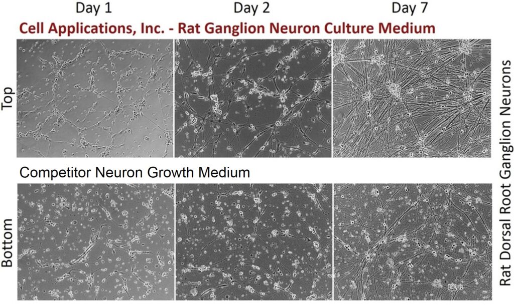
| Bioassay | Cells plate on Neuron Coating Solution II-coated surface, Arborize to form neurite network in Culture Medium | |
| Cryovial | 1,000,000 or 500,000 RDRGN in Ser-Fr Frzng Med (042-50) | |
| Kit | Cryovial RDRGN, Ctng Soln II, Pltng Med, Cultr Med Kit | |
| Doublings | N/A – Neurons don’t proliferate in vitro | |
| Applications | Laboratory research use only (RUO). Not for human, clinical, diagnostic or veterinary use. |
Resources
FAQs
Need More Help?
Visit our comprehensive FAQ page for detailed answers to common questions.
Need More Help?
Visit our comprehensive FAQ page for detailed answers to common questions.
Primary Cell FAQs Powered by Bioz