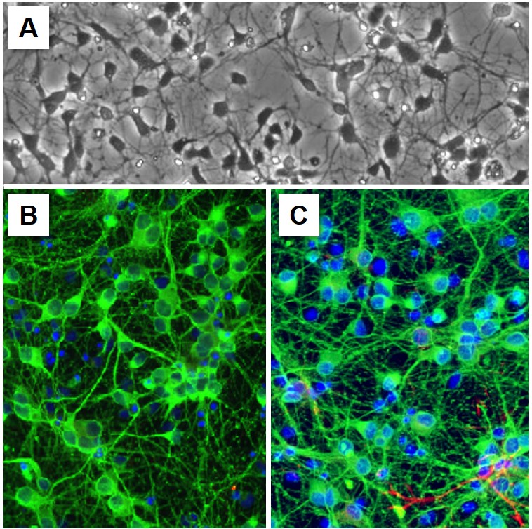
Rat Hippocampal Neurons: RHiN
Rat Hippocampal Neurons (RHiN) are a primary cell model derived from the embryonic rat hippocampus.
Description
- Neurophysiology and Plasticity: Researchers utilize these cells to investigate the cellular mechanisms of synaptic plasticity, which is the biological basis of learning and memory.
- Pathology and Disease: The hippocampus is a major site of investigation for neurodegenerative and neurological disorders. RHiN are used to model the cellular hallmarks of conditions such as Alzheimer’s disease — where hippocampal vulnerability is well-documented — as well as epilepsy, encephalitis, and the effects of hypoxia (oxygen starvation).
- Drug Discovery: By investigating how various substances affect neuronal survival, signal transduction, or protein aggregation, scientists can better understand disease mechanisms and accelerate the development of new therapeutics in medicine.
For researchers conducting a literature search regarding these cells, they are often categorized under specific MeSH (Medical Subject Headings) terms related to neural development and neurobiology. Utilizing standardized, high-quality primary cell lines ensures that the data obtained regarding neuronal health and signaling is both reproducible and physiologically relevant. By bridging the gap between molecular observation and clinical application, RHiN remain a vital tool for advancing our understanding of the brain’s complex circuitry. The study of rat hippocampus-derived neurons is helpful for deciphering the mechanisms underlying complex cognitive functions. As detailed in specialized research (e.g., doi.org/10.1089/adt.2017.797), the ability to model the neuron in a controlled environment allows for targeted investigations into the pathology of the brain, ultimately providing the data necessary to foster advancements in neurological health.
Details
| Tissue | Normal healthy rat hippocampus | |
|---|---|---|
| QC | No bacteria, yeast, fungi, mycoplasma | |
| Character | Positive for β-III Tubulin | |
| Bioassay | Plate on Poly-D-Lysine coated surface, arborize to form neurite network in Culture Med | |
| Cryovial | 1M RHiN cryopreserved after isolation in Serum-Free Freezing Med (042-50) | |
| Cryo Kit | RHiN (R886N-10), Cltr Med (R817-100) Ctng Soln I (027-05), Pltng Med (R886P-10) | |
| Doublings | N/A – Neurons don’t proliferate in vitro | |
| Applications | Laboratory research use only (RUO). Not for human, clinical, diagnostic or veterinary use. |
Resources
FAQs
Need More Help?
Visit our comprehensive FAQ page for detailed answers to common questions.
Need More Help?
Visit our comprehensive FAQ page for detailed answers to common questions.
Primary Cell FAQs Powered by Bioz