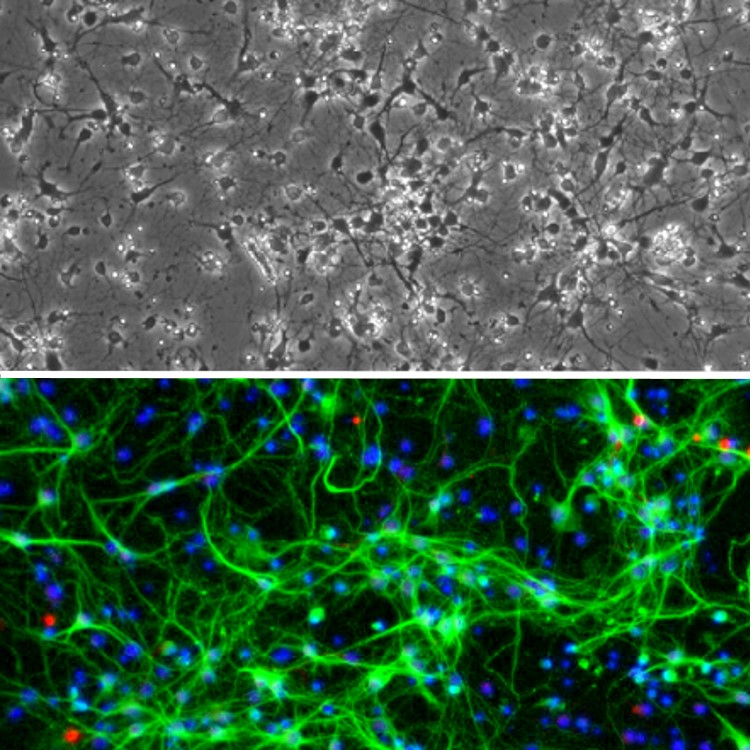
Cryopreserved Rat Hindbrain Neurons (RHbN) revived and cultured for 8 days (top), stained (bottom) for β III-Tubulin (green), GFAP (red), and DAPI (blue).
Rat Hindbrain Neurons: RHbN
Rat Hindbrain Neurons (RHbN) are primary neurons derived from the hindbrain of normal embryonic rats.
Description
Rat Hindbrain Neurons (RHbN) are primary neurons derived from the hindbrain of normal embryonic rats (commonly at gestation day 15) using standardized isolation and culture methods. RHbN develop neuritic arborization and form a complex neurite network within about a week under recommended culture conditions, and they show neuronal identity markers such as βIII-Tubulin. As a brain region encompassing structures such as the pons and medulla (and closely associated circuitry with cerebellar-related coordination), hindbrain cell types contribute to vital sensorimotor and autonomic control functions. This includes coordination and equilibrium maintenance, as well as regulation of breathing, swallowing, muscle tone, and arousal-related patterns. When hindbrain structure or function is disrupted by malformations, vascular disorders, atrophies, or trauma, these neural control roles can be impaired, producing clinical features with varying severity — making RHbN a useful system for dissecting mechanisms of physiopathology and for supporting neuronal drug discovery and therapy development.
RHbN cultures are valuable for probing neuronal activity and how specific stimuli shape firing rate and downstream network-like responses. Depending on the experimental design, studies can assess neuronal activity in terms of stimulus-dependent changes in individual neurons and coordinated responses across a neurite network, supporting systems neuroscience investigations into how neuronal activity emerges from cell type–specific properties. RHbN can also be used to explore neurotransmitter and monoaminergic phenotypes relevant to hindbrain function, including gabaergic and catecholamine-associated biology. These approaches connect cellular neuronal activity to brain research questions spanning brain region–specific function and behavior-linked outputs such as ingestive behavior and locomotor activity, through the control roles of brainstem circuits.
Because neuronal systems can respond differently across species, RHbN provide a rat brain tissue–derived platform that complements findings from mouse brain models while supporting cross-species interpretation of neuronal activity. In this context, RHbN support study of hindbrain-associated autonomic pathways and brainstem–cerebellar loops, as well as relevant monoaminergic brainstem nuclei (rather than basal ganglia or midbrain reward circuitry). For example, hindbrain models can be used to investigate catecholamine-associated signaling systems originating in brainstem regions such as the locus coeruleus and other noradrenergic or adrenergic cell groups in the medulla, alongside serotonergic inputs via raphe-related brainstem circuitry. Overall, Rat Hindbrain Neurons (RHbN) offer a controlled mammalian-relevant cell type model for characterizing neuronal activity, stimulus-driven responses, and disease-linked dysfunction in a tractable in vitro format suitable for toxicity testing, immunostaining, live cell imaging, co-culture experiments, electrophysiology, and related neuroscience and neuroscience-focused drug evaluation studies.
Rat Hindbrain Neurons (RHbN) are derived from hindbrains of normal embryonic rat (gestation day 15) by standardized methods. When cultured under the recommended conditions, RHbN arborize and form complex neurite network in one week. RHbN Stain positive for β III-Tubulin.
The hindbrain includes the cerebellum, the pons, and the medulla. These parts together control and support various vital bodily processes such as breathing, swallowing, blood circulation, muscle tone, sleep and arousal patterns, coordination, equilibrium maintenance, and more. Malformation, vascular disorders, atrophies, and traumas at hindbrain strike its proper structure and functions, leading to the manifestation of a variety of clinical features of different severity. Studying hindbrain neurons in those disease conditions will allow a better understanding of the disease mechanisms, physiopathologies, and advancement in drug development and therapies. Cultures of hindbrain neurons can be applied for a variety of experiments including toxicity test, immunostaining, live cell imaging, co-culturing, electrophysiology, and more.
Details
| Tissue | Normal healthy rat hindbrain | |
|---|---|---|
| QC | No bacteria, yeast, fungi, mycoplasma, virus | |
| Character | Positive for β-III Tubulin | |
| Bioassay | Plate on Poly-D-Lysine coated surface, arborize to form neurite network in Culture Med | |
| Cryovial | 1,000,000 RHbN in Ser-Fr Frzng Med (042-50) | |
| Kit | Cryovial frozen RHbN, Ctng Soln I (027-05), Plating Med (R817P-10), Cultr Med (R817-100) | |
| Doublings | N/A – Neurons don’t proliferate in vitro | |
| Applications | Laboratory research use only (RUO). Not for human, clinical, diagnostic or veterinary use. |
Resources
FAQs
Need More Help?
Visit our comprehensive FAQ page for detailed answers to common questions.
Need More Help?
Visit our comprehensive FAQ page for detailed answers to common questions.
Primary Cell FAQs