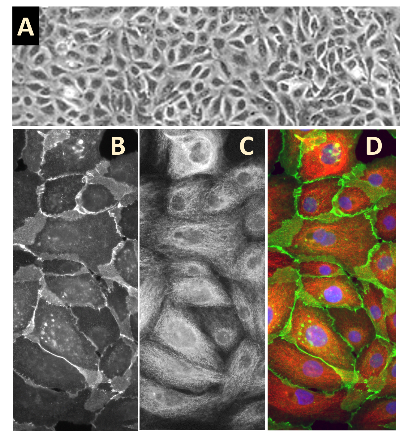
Human Mammary Epithelial Cells: HMEpC
Human Mammary Epithelial Cells (HMEpC) are primary epithelial cells derived from the complex tubuloalveolar network of the human mammary gland.
Description
Human Mammary Epithelial Cells (HMEpC) are primary cells derived from the complex tubuloalveolar network of the human mammary gland. Within the native breast architecture, the mammary epithelial cell population is organized into a bilayered structure consisting of an inner layer of secretory luminal epithelial cells and an outer layer of contractile basal myoepithelial cells. Together, these subsets regulate the structural homeostasis, cyclical remodeling, and specialized lactogenic functions of the tissue.
As primary mammary epithelial cells, HMEpC possess a finite lifespan and limited self-renewal capacity in vitro, reflecting tight physiological constraints that contrast with immortalized or transformed lines. Historically, the continuous expansion of normal human mammary epithelial cells was heavily restricted by rapid stromal fibroblast overgrowth. To address this, specialized serum-free media formulations are deployed; Bovine Pituitary Extract (BPE) is one commonly used supplement to support epithelial outgrowth, though modern defined formulations increasingly seek to replace or standardize such extracts. Rather than rendering transformation impossible, these selective culture conditions significantly reduce stromal contamination and permit the robust expansion of non-transformed epithelial isolates. However, because primary culture can still select for rare cellular variants and promote subtle genetic or epigenetic modifications over extended passaging, experimental windows must be carefully monitored to maintain translational validity. Culturing authentic primary mammary epithelial cells provides a crucial, non-transformed biological baseline to examine normal epithelial function, lineage tracking, and the earliest molecular changes that initiate malignancy.
Normal human mammary epithelial cells serve as a vital experimental control and mechanistic model system in peer-reviewed literature, particularly across molecular oncology workflows designed to deconstruct the pathways of breast tumor initiation, progression, and metastasis:
-
Breast Cancer Genesis and Stemness Models: A primary application of the human mammary epithelial cell is defining the boundaries between normal tissue homeostasis and malignant transformation. Researchers utilize HMEpC to investigate the characterization and behavior of tissue-resident stem cells and committed progenitors. Because mammary epithelial progenitor hierarchies remain an active area of research—featuring evolving, distinct lineage models and differing surface marker definitions between human and mouse tissues—HMEpC provide an essential human platform for testing open lineage theories. By establishing how tumor suppressors maintain genetic stability during normal differentiation, investigators can trace how the loss of these checkpoints allows primitive stem cells to initiate breast cancer. For example, investigators leverage HMEpC to dissect oncogenic signaling, showing that the Myc oncogene actively contributes to malignant transformation when operating in cooperative contexts alongside complementary genetic lesions (such as telomerase activation, oncogenic RAS, or p53/RB loss).
-
Tumor-Permissive Microenvironments and Exosome Signaling: In the context of breast cancer progression, normal mammary epithelia are under continuous biochemical influence from surrounding stromal and malignant elements. HMEpC are frequently deployed to investigate how exosomes and extracellular vesicles secreted by active breast cancer cells manipulate healthy, normal mammary epithelium, remodeling the local tissue into a tumor-permissive niche. Conversely, HMEpC act as a vital, non-malignant control system in drug discovery workflows, such as screening the targeted anti-tumor properties of novel compounds like cannabinoids or evaluating the therapeutic margin of stem cell microenvironment modulators.
-
Biomarker Classification and Therapeutic Target Identification: Detailed characterization of HMEpC surface properties has advanced diagnostic profiling. Studies demonstrate that the differential expression of complex cell-surface glycoproteins allows researchers to classify human breast cells into normal, benign, malignant, basal, and luminal groups, though the specific diagnostic marker panels utilized differ across individual reports. Furthermore, comparative expression profiling between HMEpC and malignant tissue has successfully identified unique enzymatic alterations—such as the upregulation of aldehyde dehydrogenase (ALDH) isoform 5A1—as a potential molecular target in specialized human breast ductal carcinoma in situ (DCIS) research models.
-
Genomic Instability, DNA Damage, and Epigenetic Regulation: To understand the triggers of breast cancer, researchers expose HMEpC to genotoxic stress to investigate cell survival networks following direct DNA damage. Primary HMEpC models have been instrumental in showing how critically shortened telomeres can initiate genomic instability, cytokinesis failure, and pathological polyploidy. Additionally, these cells are integrated into epigenetic screening assays; for instance, specific preclinical reports pairing HMEpC with human dermal fibroblasts have demonstrated that the dietary polyphenol resveratrol can attenuate or inhibit the mono-ubiquitination of histone H2B, representing a context-dependent chromatin modification observed in particular experimental systems.
-
Extracellular Matrix (ECM) Protection and Metastatic Invasiveness: In a healthy mammary gland, normal epithelium expresses a tightly regulated profile of protective proteins that shield the surrounding extracellular matrix from degradation. In metastatic breast cancer, the loss of these protective barriers is strongly associated with heightened tissue invasion. Investigators use HMEpC to demonstrate that the loss of Maspin expression correlates with unrestricted ECM degradation and heightened metastatic potential in specific models. Similarly, a reduction in extracellular superoxide dismutase (EcSOD) expression is reported to disrupt local ECM integrity and promote an invasive cellular phenotype. To counteract aggressive epithelial proliferation in vitro, preclinical and context-specific experimental studies have explored combining targeted anti-growth factor treatments with localized, low-dose phototherapy (UV-B) to evaluate synergistic growth arrest.
- Investigate the role of exosomes secreted by cancer cells in formation of tumor permissive microenvironment through manipulation of normal mammary epithelium
- Serve as control in a study investigating antitumor properties of cannabinoids and stem cell microenvironment
- Determine that differential expression of glycoproteins allows classification of human breast cells into normal, benign, malignant, basal, and luminal groups
- Identify ALDH isoform 5A1 as a potential target for treatment of human breast ductal carcinoma
- Determine that combination of an anti-growth factor treatment using phototherapy (UV-B) is more effective
- Investigate cell survival following DNA damage
- Show the important roles of tumor suppressors in mammary epithelial differentiation
- Explain protection of extracellular matrix from degradation in normal mammary epithelia,
- Show that Maspin loss in metastatic cancer leads to unrestricted ECM degradation, contributing to metastasis, and that loss of EcSOD expression also promotes invasiveness by disrupting ECM
- Investigate the role of shortened telomeres in initiation of genomic instability, cytokinesis failure and polyploidy
- Elucidate the role of Myc in malignancy by studying its ability to transform primary epithelial cells
- Demonstrate, along with Human dermal Fibroblasts, also from Cell Applications, Inc., that resveratrol inhibits mono-ubiquitination of histone H2B
Details
| Tissue | Normal healthy human mammary glands | |
|---|---|---|
| QC | No bacteria, yeast, fungi, mycoplasma, virus | |
| Bioassay | Attach, spread, proliferate in Growth med | |
| Cryovial | 500,000 HMEpC (5th passage) frozen in Basal Medium w/ 10% FBS, 10% DMSO | |
| Kit | Cryovial frozen HMEpC(830-05a), Growth Med (815-500), Subculture Rgnt Kit (090K) | |
| Proliferating | Shipped in Gr Med, 6th psg (flasks or plates) | |
| Doublings: | At least 16 | |
| Applications | Laboratory research use only (RUO). Not for human, clinical, diagnostic or veterinary use. |
Resources
FAQs
Need More Help?
Visit our comprehensive FAQ page for detailed answers to common questions.
Need More Help?
Visit our comprehensive FAQ page for detailed answers to common questions.
Primary Cell FAQs Powered by Bioz