Skin
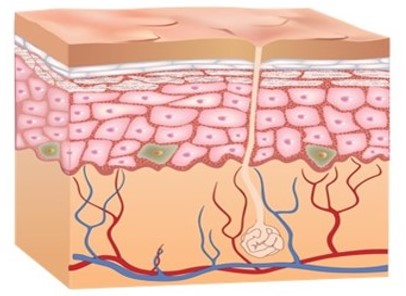
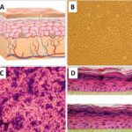
Skin is the largest organ of the human body, serving as a dynamic, stratified barrier that protects against mechanical trauma, ultraviolet (UV) radiation, fluid loss, and microbial invasion. The functional integrity of the cutaneous system relies on a continuous reciprocal dialogue between the avascular, highly cellular outer epidermis and the underlying, fibrovascular dermis. This architectural network regulates systemic thermoregulation, executes wound-healing cascades, and maintains a highly specialized localized immune environment.
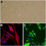
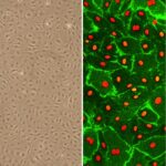
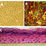
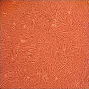
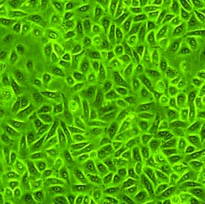
Because the structural hierarchy of intact skin is difficult to fully capture in isolated monolayer screens, contemporary dermatological, cosmetic, and toxicological research utilizes an integrated matrix of primary cells alongside complex 3D Skin Models (tissue-engineered organotypic skin equivalents). The core of this testing framework relies on Human Epidermal Keratinocytes (HEK), Human Dermal Fibroblasts (HDF), and Human Epidermal Melanocytes (HEM).
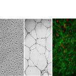
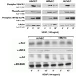
By analyzing these baseline cell lots alongside vascular elements like Human Dermal Microvascular Endothelial Cells (CADMEC/HMVEC)—alternatively designated in the literature as HMVEC-D or simply dermal HMVEC—specialized follicular structures like Human Hair Follicle Dermal Papilla Cells (HFDPC), and complementary mammalian models—such as Rat Dermal Fibroblasts (RDF) and Rat Epidermal Keratinocytes (REK)—investigators can model psoriasis, study melanoma progression, execute non-animal safety testing, and map the cellular mechanics of cutaneous aging.
However, explicit interspecies differences in skin structure, barrier thickness, lipid composition, and localized immune cell profiles mean that animal-derived cutaneous cells do not fully replicate human skin physiology, limiting direct translation for transdermal drug delivery and inflammatory disease modeling.