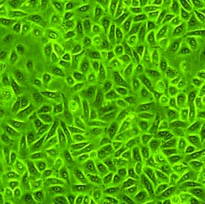
Rat Epidermal Keratinocytes: REK. Keratin producing cells isolated from skin tissue.
Rat Epidermal Keratinocytes: REK
Rat Epidermal Keratinocytes (HEK) are keratinocyte cells from the rat epidermis, the outermost layer of the skin
Description
Rat Epidermal Keratinocytes (REK) provide an in vitro model system that can be used to study aspects of epithelial function and disease, particularly when investigating pathways relevant to skin biology, toxicology, and cellular stress responses.
In vivo, the epidermis represents the outermost protective barrier of the skin, functioning as a stratified squamous epithelium where keratinocytes serve as the predominant cell type. Organized across a basal layer and outer strata, living epidermal cells maintain tissue homeostasis, protect against mechanical and environmental insults, and interact with other resident populations such as melanocytes. These cells contribute to cutaneous protective functions, barrier integrity, and local immunity by synthesizing an abundant protein framework rich in keratin, expressing various adhesion molecules, and can release cytokines in response to microenvironmental changes, inflammation, or injury.
In experimental research, rat primary cells such as Rat Epidermal Keratinocytes (REK) — alongside related models like mouse epidermal cells and human keratinocytes — can be used to investigate mechanisms relevant to keratinocyte proliferation, cell cycle regulation, cell death, and cancer development. For instance, studies have utilized REK to examine how transforming growth factor beta 1 (TGFβ1) treatment is reported to be associated with c-Myc expression and proliferation. Furthermore, these models can be used to examine how keratinocyte sodium channel expression and activity may affect ATP release and neuron-linked signaling, as well as how factors like ionizing radiation have been studied in relation to laminin 332 dynamics and can model aspects of delayed wound healing.
Maintaining experimental integrity requires rigorous handling practices, including proper storage in cryogenic vials and routine screening against mycoplasma, fungi, and other contaminants to ensure reliable results when evaluating keratinocyte behavior and genetic perturbation experiments using cells.
Rat Epidermal Keratinocytes (REK) from Cell Applications, Inc. provide a good model system to study many aspects of epithelial function and disease, particularly those related to skin biology and toxicology.
- It was shown that TGFβ1 treatment decreased c-Myc mRNA in REK which, taken together with the TGFβ1-induced growth arrest exhibited by keratinocytes and opposite results obtained in other cells, suggests a correlation between c-Myc mRNA expression and the mitogenic response.
- Additionally, REK were used to demonstrate that pathological increases in keratinocyte sodium channels Nav1.1, Nav1.6, and Nav1.8 expression may contribute to pain by increasing epidermal ATP release, resulting in excessive activation of P2X receptors on primary sensory axons.
- REK were also used to demonstrate that laminin 332 deposition is inhibited by ionizing radiation and, in combination with slower keratinocyte migration, can contribute to the delayed wound healing of irradiated skin.
Details
| Tissue | Normal healthy rat skin | |
|---|---|---|
| QC | No bacteria, yeast, fungi, mycoplasma | |
| Bioassay | Attach, spread, proliferate in Growth Med | |
| Cryovial | 500,000 REK (primary culture) frozen in Basal Medium w/ 10% FBS, 10% DMSO | |
| Kit | Cryovial frozen REK (R102-05n), Growth Medium (R131-250) | |
| Proliferating | Shipped in Gr Med, 1st psg (flasks or plates) | |
| Doublings | REK do not expand and cannot be subcutured. Thaw & plate REK directly to assay format. REK will undergo only 1 or 2 doublings. | |
| Applications | Laboratory research use only (RUO). Not for human, clinical, diagnostic or veterinary use. |
Resources
FAQs
Need More Help?
Visit our comprehensive FAQ page for detailed answers to common questions.
Need More Help?
Visit our comprehensive FAQ page for detailed answers to common questions.
Primary Cell FAQs Powered by Bioz