3D Airway Model
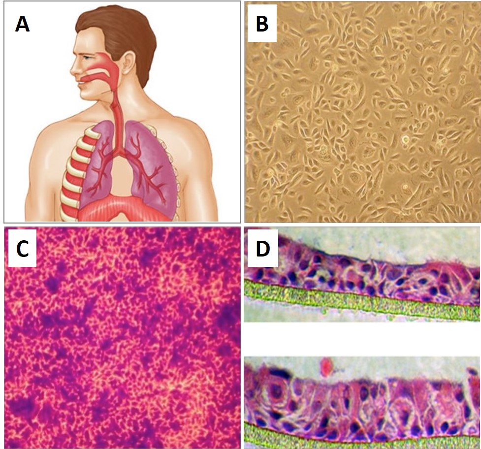
The 3D Airway Model is a three-dimensional tissue-engineered system composed of primary Human Bronchial Epithelial Cells (HBEpC).
Description
Under these optimized ALI culture conditions, the cells polarize and mature into a differentiated pseudostriated epithelium containing ciliated cells, mucus-secreting goblet cells, and regenerative basal cells. While this system reproduces key epithelial barrier features, investigators must note that isolated ALI HBEpC cultures lack full multicellular airway complexity — such as resident immune cell populations, functional vasculature, submucosal glands, airway smooth muscle, and an intact native extracellular matrix — unless specifically co-cultured or bioengineered. The synchronized beating of the ciliated cells, combined with the endogenous production of protective mucus layer components, establishes an active mucociliary transport mechanism in vitro. However, functional endpoints for mucociliary clearance in vitro are typically assessed indirectly via cilia beat frequency, particle transport assays, or mucus production metrics, and these do not fully reproduce complex, whole-organ clearance dynamics.
Differentiated 3D Airway Models provide an indispensable experimental and translational framework for exploring respiratory pathology, tissue remodeling, and advanced bioengineering workflows:
-
Pulmonary Disease Modeling and Stress Responses: The model facilitates the detailed in vitro examination of epithelial function, ciliary dyskinesia, and targeted signaling alterations under pathological conditions. Researchers utilize this platform to model chronic airway diseases, such as asthma, chronic obstructive pulmonary disease (COPD), and cystic fibrosis, by exposing the mature barrier to specific inflammatory cytokines or allergens. While modeling complex chronic pathologies in vitro can reproduce certain cellular phenotypes, it cannot fully recapitulate systemic, multicellular, and temporal disease features without additional components or long-term, multi-cell-type culture systems. The accessible mucosal surface allows investigators to quantify cellular responses to mechanical stress, oxidative injury, viral/bacterial airway infections, and airborne toxicants like cigarette smoke, accelerating the discovery of novel therapeutic interventions for acute lung injuries.
-
Digital Imaging, Mesh Generation, and Computational Fluid Dynamics: To translate these cellular findings into macro-level physiological frameworks, researchers combine biological data with advanced digital imaging techniques. High-resolution micro-CT or MRI scans of the respiratory tree generate detailed volumetric datasets. From this raw data, specialized segmentation software extracts the complex luminal boundaries to construct a precise digital mesh. This computational 3D model can then be subjected to computational fluid dynamics (CFD) testing to map airflow resistance, shear stress profiles, and aerosolized drug deposition patterns. However, because matching in vitro transport rates to in vivo deposition requires careful scaling and validation, researchers must account for these complex parameters rather than assuming a straightforward, one-to-one mapping between the culture dish and computational modeling software.
-
3D Printing and Surgical Planning Interventions: Beyond computational software, the integration of bio-anatomical data bridges the gap into clinical medicine via additive manufacturing. By exporting a patient-specific mesh to high-fidelity 3D printing systems, engineers can manufacture a physical, tactile 3D printed model that replicates the exact dimensions of a patient’s airway anatomy. These physical models provide an invaluable resource for a thoracic surgeon preparing for complex reconstructive surgeries, tracheal stenoses, or tumor resections. Having open access to a highly accurate 3D printed model allows an expert clinical team to perform preoperative trial fittings of custom stents, anticipate challenging structural variations, and drastically increase surgical confidence prior to entering the operating theatre.
Details
FAQs
Need More Help?
Visit our comprehensive FAQ page for detailed answers to common questions.
Need More Help?
Visit our comprehensive FAQ page for detailed answers to common questions.
Primary Cell FAQs Powered by Bioz