Human Bronchial Epithelial Cells: Asthma: HBEpC-AS
Human Bronchial Epithelial Cells: Asthma (HBEpC-AS) are primary epithelial cells isolated from the surface of human bronchi obtained from donors clinically diagnosed with asthma.
Description
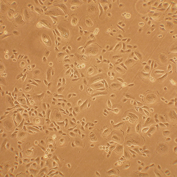
Human Bronchial Epithelial Cells: Asthma (HBEpC-AS) are primary epithelial cells isolated from the surface of human bronchi obtained from donors clinically diagnosed with asthma. As a primary human bronchial epithelial cell type rather than an immortalized cell line, HBEpC-AS may retain an ex vivo disease-associated memory of the chronic airway inflammation and remodeling characteristic of asthmatic patient lungs. The retention and persistence of these in vivo asthma-specific signatures ex vivo are conditional and variable, depending on factors such as isolation timing, prior treatment regimens (especially exposure to corticosteroids), cell selection methods, and the number of in vitro passages. For definitive authentication, these cells display a classic cobblestone morphology consistent with epithelial origin and test positive for the specific epithelial cell marker cytokeratin 18, making them a translationally relevant model when carefully controlled.
In the human body, these airway epithelial cells form the continuous mucosal lining of the conducting airways within the human bronchi. This specialized airway epithelium acts as the primary physical and immunological barrier separating the underlying lung tissue from the external environment, where it is constantly exposed to inspired oxygen and inhaled foreign particles. In asthmatic patients, this barrier is often compromised, showing structural alterations and increased permeability; however, this barrier dysfunction is not universal, and its degree varies significantly by patient phenotype and clinical control. When present, this altered barrier places the airway epithelia at the epicenter of structural tissue remodeling, allowing for close interaction with localized immune cells and underlying airway smooth muscle. Consequently, these primary cells are highly valuable for studying localized respiratory disease dynamics, particularly how mucosal surfaces function in individuals with a history of frequent asthma symptoms or recurrent atopic asthma flare-ups.
In a healthy respiratory tract, the primary function of the bronchial epithelium is to orchestrate mechanical clearance via ciliary beating, maintain barrier integrity, and initiate a balanced innate immune response against environmental insults. In asthmatics, however, the airway epithelium often shows a dysfunctional, pro-inflammatory phenotype under certain conditions. When exposed to common allergy components or environmental asthma triggers, the epithelial cells can secrete an altered profile of cytokines, chemokines, and growth factors. This dysregulated signaling recruits and activates surrounding immune cells, contributing to localized airway inflammation, driving mucus hypersecretion, and inducing hyperreactivity in the adjacent airway smooth muscle. This structural and immunological cross-talk can undermine overall lung function and play a role in provoking an asthma attack or acute asthma exacerbation, which narrows the air passageways.
In laboratory settings, HBEpC-AS serve as an important model in respiratory research and can be used in targeted drug discovery cell screening to complement high-throughput work done in cell lines. When grown on specialized culture inserts and transitioned to an air-liquid interface (ALI), these cells differentiate into a pseudostratified, ciliated tissue configuration that models the 3D architecture of the human respiratory tract. Investigators utilize this system to examine the molecular pathways driving allergic asthma, study chronic comorbidities like chronic rhinosinusitis, and evaluate the specific cellular mechanisms behind an asthma attack. Furthermore, healthcare organizations—such as the American Lung Association—emphasize the need for translational models to improve asthma care, advance general asthma management, and refine clinical asthma treatment protocols. However, linking in vitro responses directly to clinical interventions—such as evaluating how an asthma action plan, specific asthma medication choices, or non-pharmacological therapies like bronchial thermoplasty impact epithelial repair—requires multiple validation steps, including robust donor replication, appropriate healthy controls, and careful clinical correlation.
Human Bronchial Epithelial Cells (HBEpC) provide an excellent model system to study all aspects of epithelial function and disease, particularly those related to airway viral infections, as well as tissue repair mechanisms, signaling changes and potential treatments relevant to lung injuries, mechanical and oxidative stress, inflammation, pulmonary diseases and smoking. When grown on inserts and provided with the liquid/air interface, HBEpC can differentiate into a pseudostriated epithelium and serve as a more physiological 3D tissue model for in vitro studies. The HBEpC shown here were cultured (L) and immunolabeled for cytokeratin 18 (R). 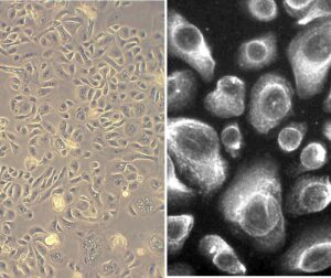
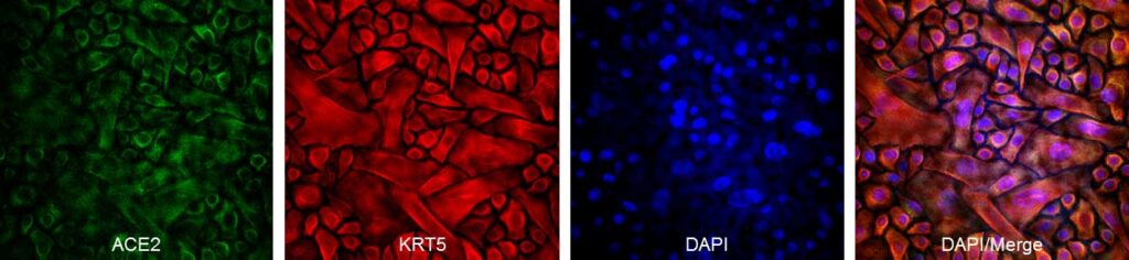
- Activation, expression and production of genes, kinases and signaling pathways by cytokines, growth factors, interleukins, binding proteins and pro-inflammatory molecules.
- Stimulation-dependent, observable changes in proliferation, bronchial epithelial permeability, crosslinking of membrane glycoproteins and cell surface adhesion molecules. Drug discovery cell screening for in vitro assay of compounds, or to extend and confirm high-throughput work done in cell lines.
- Clinical focused discoveries leveraging HBEpC include therapeutics to suppress tumor gene transcription, apoptosis, inflammation, auto-immune disease and viral infection, while enhancing cell protection, repair and lifespan.
Details
| Tissue | Human bronchial epithelium: Asthma | |
|---|---|---|
| QC | No bacteria, yeast, fungi, mycoplasma, virus | |
| Bioassay | Attach, spread, proliferate in Growth Med | |
| Cryovial | 500,000 HBEpC-AS (1st passage) frozen in Basal Med w/ 10% FBS, 10% DMSO | |
| Kit | Cryovial frozen HBEpC-AS (502AS-05a), Gr Med (511-500), Subculture Rgnt Kit (090K) | |
| Proliferating | Shipped in Gr Med, 3rd psg (flasks or plates) | |
| Doublings | At least 16 | |
| Applications | Laboratory research use only (RUO). Not for human, clinical, diagnostic or veterinary use. |
Resources
FAQs
Need More Help?
Visit our comprehensive FAQ page for detailed answers to common questions.
Need More Help?
Visit our comprehensive FAQ page for detailed answers to common questions.
Primary Cell FAQs