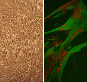
Human Skeletal Muscle Cells, HSkMC (L). Fused HSkMC (fetal) immunolabeled for desmin (green), nuclei visualized with PI (red) (R).
Human Skeletal Muscle Cells: HSkMC
Human Skeletal Muscle Cells (HSkMC) are specialized myogenic cells responsible for the structure and function of voluntary muscle tissue.
Description
Human Skeletal Muscle Cells (HSkMC) are specialized myogenic cells responsible for the structure and function of voluntary muscle tissue. In tissue biology and experimental workflows, the nomenclature of this cell type shifts depending on differentiation and maturation status. The term myoblasts refers to the proliferative, mononucleated cells that act as myogenic progenitors. In contrast, skeletal myocytes or myotubes denote the post-mitotic, fused, and differentiated state. While satellite cells (or skeletal muscle satellite cells) serve as the native, resident stem and progenitor pool in living tissue, investigators should note that primary cultures typically consist of a dynamic mix of these activated progenitors and proliferating myoblasts rather than a pure, quiescent satellite cell population. To support their expansion in vitro without triggering premature or spontaneous differentiation, they must be maintained using a highly optimized, nutrient-rich Human Skeletal Muscle Growth Medium.
In the human body, skeletal muscle is the largest organ by mass, comprising ~40% of body mass in adults. This tissue is distributed globally across the sub-cutaneous skeleton to facilitate voluntary locomotion, maintain postural stability, and support respiratory mechanics. Within this highly vascularized and innervated microenvironment, HSkMC are organized into parallel bundles of multinucleated fibers. While early alpha cytology and classic developmental studies mapped these structures in donor limbs, modern research frequently investigates how HSkMC cross-talk with circulating blood cells and mononuclear cells from the peripheral blood. For instance, during injury, a wave of immune entities — including monocytes, T cells, and natural killer (NK) cells — infiltrates the tissue to clear cellular debris and release paracrine signals that coordinate local muscle regeneration.
The primary biological function of mature HSkMC is to undergo synchronized contraction to generate mechanical force, while simultaneously serving as a principal hub for systemic metabolic homeostasis. During skeletal muscle differentiation, mononucleated myoblasts align and fuse to form elongated, multinucleated myotubes that synthesize organized sarcomeric myofilaments. Aside from motor function, these cells represent the primary site for insulin-stimulated glucose disposal. They integrate complex endocrine signals to regulate glucose kinetics, activating downstream networks like the PI3K/Akt signaling pathway to trigger the translocation of glucose transporter 4 (GLUT4) to the cell membrane. Furthermore, they function as an active metabolic rheostat during nutrient deprivation, altering their gene expression profiles to manage organic acid metabolism and fuel selection under energetic stress.
In laboratory settings, primary HSkMC provide a translationally rigorous human background model for mapping muscle development, investigating metabolic diseases like type 2 diabetes, and evaluating safety pharmacology. Researchers routinely expose HSkMC to free fatty acids or inflammatory cytokines to induce state-specific insulin resistance in vitro, allowing for the precise screening of anti-diabetic compounds. Publications in journals like Cell Res highlight their utility in characterizing statin-induced myotoxicity, modeling rare genetic myopathies, and screening neuromuscular therapeutics.
Beyond mature muscle physiology, HSkMC serve as an essential somatic baseline control group in regenerative medicine. They are widely deployed to evaluate the efficiency and safety of directing pluripotent stem cells — such as embryonic stem cells and induced pluripotent stem cells — along the line of myogenic differentiation. Furthermore, they are used to test the gene transfection efficiencies of advanced non-viral vectors designed to treat muscular dystrophies.
Understanding Cross-Talk in Humanized Models
To evaluate how human muscle cells interact with a humanized immune system in vivo, advanced translational workflows utilize highly immunodeficient mouse strains reconstituted with human peripheral blood mononuclear cells (huPBMC) or human hematopoietic stem cells (HSCs). These platforms allow researchers to evaluate human cell-to-cell dynamics within a living system:
-
huPBMC Humanized Model: Created by transferring mature human peripheral blood mononuclear cells (PBMC/HMNC-PB) into immunodeficient mice. This system is highly valuable for short-term studies analyzing the direct activation and homing of mature human T cells, B cells, or monocytes. However, researchers must account for severe Graft-versus-Host Disease (GvHD) limits inherent to huPBMC models, which typically constrains the experimental window to a few weeks.
-
Hematopoietic Stem Cells (HSCs): Isolated from bone marrow, umbilical cord blood, or mobilized peripheral blood. When human hematopoietic stem cells are engrafted into newborn mice, they give rise to a complete, long-term human immune system, producing continuous generations of diverse hematopoietic cells, including T cells, B cells, natural killer (NK) cells, and myeloid progenitors.
-
Established Strain Terminology: To minimize mouse-derived immune interference, optimize human cell engraftment, and delay GvHD, these models rely on highly specialized immunodeficient backgrounds. Common platforms include NSG (NOD.Cg-Prkdcscid Il2rgtm1Wjl/SzJ), NOG, and BRG mice. Advanced variations incorporate targeted murine major histocompatibility complex (MHC) or HLA-knockout variants, allowing investigators to precisely analyze human leukocyte kinetics and targeted antitumor activity without confounding background graft rejection.
- Serve as a differentiated control in a study of developmental regulator genes in hESC
- Characterize statin-induced gene expression changes and demonstrate cytotoxic effect of statins in skeletal muscle cells
- Identify molecular mechanisms of mitochondrial myopathy and sideroblastic anemia resulting from a missense mutation in the PUS1 gene
- Investigate chemokine-like factor expression in the idiopathic inflammatory myopathies
- Demonstrate that fasting activates AceCS2 gene expression by inducing KLF15 transcription factor
- Characterize human FGFR3-positive sarcoma-initiating stem cells
- Identify trans-Golgi network proteins and Notch and Hedgehog pathways as putative targets for rhabdomyosarcoma therapy
- Show that riluzole muscle relaxant effects are mediated by inhibition of INa and stimulation of BKC-channel activity
- Develop nitric esters that combine the pharmacological functions of NO and muscle relaxation properties for treatment of muscular diseases
- Develop biodegradable polymer-based transgene delivery vectors for muscular dystrophy treatment
- Design optimal coating for orthopedic metallic implants
Details
| Tissue | Normal healthy human limb skeletal muscle | |
|---|---|---|
| QC | No bacteria, yeast, fungi, mycoplasma, virus | |
| Character | Multinucleated myotubes form after culture in fusion medium | |
| Bioassay | Attach, spread, proliferate in Growth Med | |
| Cryovial | 500,000 HSkMC (2nd passage) frozen in Basal Medium w/ 10% FBS, 10% DMSO | |
| Kit | Cryovial frozen HSkMC (150-05), Growth Medium (151-500), Subcltr Rgnt Kit (090K) | |
| Proliferating | Shipped in Tsfr Med, psg 3, flasks or plates | |
| Doublings | At least 15 | |
| Applications | Laboratory research use only (RUO). Not for human, clinical, diagnostic or veterinary use. |
Resources
FAQs
Need More Help?
Visit our comprehensive FAQ page for detailed answers to common questions.
Need More Help?
Visit our comprehensive FAQ page for detailed answers to common questions.
Primary Cell FAQs Powered by Bioz