Joint
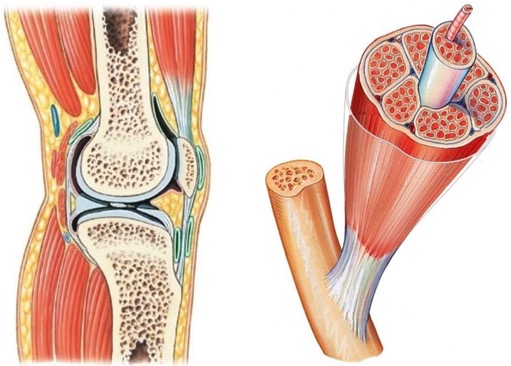
Diarthrodial (synovial) joints are highly evolved mechanical structures designed to facilitate smooth, low-friction locomotion while distributing heavy compressive loads across the skeletal framework. Rather than acting as a simple structural hinge, a functioning joint relies on a tightly coordinated tissue dialogue between the avascular, largely non-innervated articular cartilage matrix and the highly vascular, specialized synovial membrane. While deeper articular cartilage zones are completely aneural, the superficial zone near the synovium and the adjacent subchondral bone regions can exhibit pain-sensitive innervation under mechanical stress or pathological degeneration.
Because accessing healthy, intact human joint tissue presents severe clinical hurdles and primary joint cells undergo rapid phenotypic drift when isolated, contemporary orthopedic and rheumatology research utilizes a specialized matrix of primary cellular platforms. The core of this testing grid consists of matrix-secreting Human Chondrocytes (HC) and immunologically active Human Fibroblast-Like Synoviocytes (HFLS).
By analyzing these baseline cell lots alongside disease-specific cohorts (Osteoarthritis and Rheumatoid Arthritis), Human Re-Differentiated Chondrocytes (HC-RD), and complementary mammalian models—such as Canine Chondrocytes (CnC)—investigators can model progressive cartilage degradation, map localized pan-synovial inflammation, evaluate biomaterial scaffold integration, and screen targeted anti-inflammatory or chondroprotective therapies.
