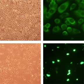
Top: Human Tracheal Epithelial Cells: HTEpC (L). HTEpC immunolabeled with an anti-CK18 antibody (R). Bottom: HTEpC (L) transfected with GFP plasmid DNA using the Cytofect™ Epithelial Cell Transfection Kit (R).
Human Tracheal Epithelial Cells: HTEpC
Human Tracheal Epithelial Cells (HTEpC) are a primary cell type isolated from the surface epithelial cells of the human trachea.
Description
Human Tracheal Epithelial Cells (HTEpC) are a primary cell type isolated from the surface epithelial cells of the human trachea. They form the essential physical and immunological barrier of the respiratory system. Unlike a transformed cell line, which may undergo genetic or phenotypic drift, HTEpC retain the physiological characteristics of the donor tissue, making them a widely used model for disease modeling.
While cytokeratin 18 (CK18) is often used for lineage identification, researchers utilize combined marker panels to distinguish between cell states. Basal progenitor cells are identified by KRT5/KRT14 and p63 expression, while differentiated luminal cells — including ciliated cells and secretory cells — express CK8/CK18. Along with human bronchial epithelial cells (HBEpC), HTEpC are among the most physiologically relevant models for studying the proximal airway, offering a high-fidelity system to investigate airway pathobiology.
In the native respiratory epithelium, these cells form a pseudostratified ciliated columnar layer. They are specialized to orchestrate mucociliary clearance, a vital innate defense where coordinated ciliary beating traps and expels inhaled particulates and pathogens.
Researchers utilize HTEpC to study the homeostasis of this barrier. By sourcing cells from distinct donor cohorts (e.g., healthy vs. asthmatic), they allow for direct comparative analysis of epithelial dysfunction and susceptibility to environmental stressors without the confounding variables associated with immortalized cell models.
Structural remodeling, particularly mucous hypersecretion and goblet cell hyperplasia, is a hallmark of asthma and COPD. HTEpC are a robust model for these investigations:
- Transcriptional Regulation: HTEpC serve as a model for investigating goblet cell differentiation. SPDEF acts as a transcriptional gateway that promotes goblet cell programs while antagonizing FOXJ1-mediated ciliated differentiation through competitive interactions and transcriptional repression.
- Environmental Stressors: These cells are used to evaluate how cigarette smoke, electronic cigarette vapor, and mechanical injury disrupt tight junctions (e.g., ZO-1 and occludin expression) and induce oxidative stress, providing insights into how epithelial integrity is compromised in chronic inflammatory states.
As the primary entry point for airborne pathogens, the tracheal epithelium is an active immunological interface.
- Viral and Bacterial Dynamics: HTEpC are a widely cited model for mapping the infection mechanics of RSV, influenza, and adenoviruses. Researchers use them to map viral entry receptors, assess replication kinetics, and monitor the secretion of pro-inflammatory cytokines like IL-8.
- Therapeutic Screening: Pharmacological studies leverage this system to screen anti-inflammatory compounds. Investigators may assess the efficacy of IL-1 receptor antagonists (e.g., anakinra) or targeted kinase inhibitors in protecting barrier integrity and attenuating hyper-inflammation.
To maximize the potential of HTEpC, investigators must carefully select their culture vessel and growth conditions:
- Submerged 2D Culture: When grown in standard plastic vessels using a specialized HTEpC growth medium, these cells proliferate as a monolayer. While this configuration allows for expansion, it results in reduced differentiation and lacks the polarized architecture and coordinated ciliary function characteristic of in vivo tissue.
- Air-Liquid Interface (ALI): For functional studies, researchers transition cells to an ALI configuration on porous inserts. Depriving the apical surface of media induces polarization, the formation of functional tight junctions, and the development of cilia. This maturation process typically requires 2–4 weeks for robust ciliary development.
Primary respiratory epithelial cells are prone to phenotypic drift and passage-dependent senescence. As cultures undergo repeated passaging, they often shift toward a more mesenchymal or undifferentiated state, which can compromise the validity of functional assays. Therefore, researchers must utilize cells at low passage numbers and monitor differentiation markers consistently to ensure experimental reproducibility. HTEpC are highly sensitive to cryopreservation, and optimized cryoprotectant protocols are essential to ensure high cell viability and post-thaw functionality.
Human Tracheal Epithelial Cells (HTEpC) provide a useful model system to study all aspects of epithelial function and disease, particularly those related to airway viral infections, as well as tissue repair mechanisms, signaling changes and potential treatments relevant to lung injuries, mechanical and oxidative stress, pulmonary diseases and smoking. Note that Cell Applications, Inc. offers HTEpC obtained from healthy donors, as well as from asthma patients.
HTEpC from Cell Applications, Inc. have been used in a study investigating the role of goblet cell hyperplasia, possibly mediated by the expression of SPDEF, to the pathogenesis of chronic pulmonary diseases.
Characterization: Morphology consistent with epithelial origin, and positive for epithelial cell marker cytokeratin 18
Details
| Tissue | Surface epithelium of normal healthy human trachea | |
|---|---|---|
| QC | No bacteria, yeast, fungi, mycoplasma, virus | |
| Bioassay | Attach, spread, proliferate in Growth Medium | |
| Cryovial | 500,000 HTEpC (1st passage) frozen in Basal Medium w/ 10% FBS, 10% DMSO | |
| Kit | Cryovial frozen HTEpC (504-05a), Growth Medium (511-500), Subcltr Rgnt Kit (090K) | |
| Proliferating | Shipped in Gr Med, 2nd psg (flasks or plates) | |
| Doublings | At least 16 | |
| Applications | Laboratory research use only (RUO). Not for human, clinical, diagnostic or veterinary use. |
Resources
FAQs
Need More Help?
Visit our comprehensive FAQ page for detailed answers to common questions.
Need More Help?
Visit our comprehensive FAQ page for detailed answers to common questions.
Primary Cell FAQs Powered by Bioz