Description
BACKGROUND Src family kinases (SFKs) consist of eight non-receptor tyrosine kinases (Src, Fyn, Yes, Lck, Lyn, Hck, Fgr and Blk) that interact with the intracellular domains of growth factor/cytokine receptors, GPCRs and integrins. Further division of the SFKs into groups A and B has been suggested based on their tissue expression profiles. Group A includes Src, Fyn, Hck, and Yes, which are expressed ubiquitously, although with various levels in different cell types. In contrast, SFK group B, consisting of Hck, Fgr, Lck, Blk, and Lyn, is expressed primarily in hematopoietic cells. Members of the Src kinase family have a very similar domain structure with a high degree of homology in the SH1 (catalytic), linker, SH2 (p-Tyr binding), SH3 (protein-protein interaction) and SH4 (membrane association) domains. c-Src, Fyn and Yes are ubiquitously expressed, although high levels of c-Src are found in platelets, neural tissue and osteoclasts. For c-Src, autophosphorylation of Tyr416 and dephosphorylation of Tyr527 is required to switch the kinase from the inactive closed formation to the active open formation.1 c-Src can be inactivated by two kinases, c-Src kinase (CSK) and CSK homologous kinase (CHK), both of which phosphorylate Tyr527 of c-Src. The activity of the Src kinase family can also be regulated by phosphatases (e.g. SHP1), binding to adaptor proteins (e.g. Cbp) and proteasomal degradation. Src kinases are key upstream mediators of both the PI 3-K and MAPK signaling pathways, and have been shown to have important roles in cell proliferation, migration and survival.2
The fyn proto-oncogene encodes a 59-kD membrane-associated protein-tyrosine kinase that is implicated in the control of cell growth. It associates with the p85 subunit of phosphatidylinositol 3-kinase and interacts with the fyn-binding protein. It plays a role in the regulation of intracellular calcium levels. It was demonstrated that Fyn is required in brain development and mature brain function with important roles in the regulation of axon growth, axon guidance, and neurite extension.3
REFERENCES
1. Homsi J et al., Expert Opin Ther Targets. 11:91-100, 2007.
2. Bradshaw, J.M.: Cell Signal 22:1175-84, 2010
3. Huber, A.B. et al: Ann. Rev. Neurosci. 26:509-63, 2003
2. Bradshaw, J.M.: Cell Signal 22:1175-84, 2010
3. Huber, A.B. et al: Ann. Rev. Neurosci. 26:509-63, 2003
Products are for research use only. They are not intended for human, animal, or diagnostic applications.
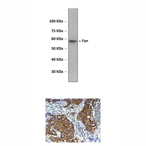
Top: Western Blot detection of Fyn proteins in NIH3T3 cell lysate using Fyn Antibody.
Bottom: This antibody also stains paraffin-embedded human breast cancer tissue in IHC.
Details
|
Cat.No.:
|
CG1165
|
|
Antigen:
|
Short peptide from N-terminal sequence of human Fyn.
|
|
Isotype:
|
Rabbit IgG
|
|
Species & predicted
species cross-
reactivity ( ):
|
Human, Mouse, Rat
|
|
Applications &
Suggested starting
dilutions:*
|
WB 1:2000
IP n/d
IHC 1:50 - 1:100
ICC 1:50 - 1:100
FACS n/d
|
|
Predicted Molecular
Weight of protein:
|
59 kDa
|
|
Specificity/Sensitivity:
|
Detects endogenous Fyn proteins without cross-reactivity with other family members.
|
|
Storage:
|
Store at -20°C, 4°C for frequent use. Avoid repeated freeze-thaw cycles.
|
*Optimal working dilutions must be determined by end user.