Product Sheet CP10234
Description
BACKGROUND Tcf proteins (Tcf1, Tcf3, Tcf4, and Lef1 in mammals) are the DNA-binding transcriptional regulators of the canonical Wnt signaling pathway, which play important role in cell fate decisions and differentiation. Although some contexts allow certain Tcfs to share a degree of functional redundancy, different Tcf family members do not always behave similarly when expressed in the same cell type. Through a highly conserved HMG domain and an amino-terminal beta-catenin interaction domain, each Tcf protein can promote transcription of downstream targets (for example, siamois and Xnr3) when Wnt-stabilized beta-catenin accumulates intracellularly. In the absence of stabilized beta-catenin, Tcf proteins have been shown to function as transcriptional repressors by interacting with corepressor proteins, such as Groucho, CtBP, and HIC-5. Interaction between Tcf3 and beta-catenin occurs at the NH2 terminus of Tcf3 and is separate from the Groucho and CtBP binding regions. Models of the wnt pathway suggest a role for Tcf3 as an unregulated scavenger of free beta-catenin. However, CBP binds and acetylates a lysine in the beta-catenin interaction domain of Tcf3, thereby lowering its affinity for beta-catenin. Some members of the Sox family of HMG box proteins also bind beta-catenin and block its binding to Tcf. In Caenorhabditis elegans, components of the mitogen-activated kinase pathway phosphorylate Tcf-bound beta-catenin so as to block nuclear localization. These studies indicate that the interaction between beta-catenin and Tcf3 is dynamic and that regulating it may play an important role in modulating wnt signaling. It was demonstrated that GSK3 and casein kinase (CK) 1epsilon both have direct but opposite effects in regulating the beta-catenin–Tcf3 interaction. Phosphorylation of Tcf3 by CKIepsilon stimulates its binding to beta-catenin, an effect reversed by GSK3. Tcf3 synergizes with CK1epsilon to inhibit beta-catenin degradation.1
Direct relationships between the biochemical properties of Tcf proteins and their physiological effects have been demonstrated by several studies expressing mutated forms of the proteins in model organisms. Three transcription factors, Nanog, Oct4, and Sox2, have been reported to form a feedforward circuit promoting pluripotent cell self-renewal in embryonic stem cells (ESC). It was shown that Tcf3 acts broadly on a genome-wide scale to reduce the levels of several promoters of self-renewal (Nanog, Tcl1, Tbx3, Esrrb) while not affecting other ESC genes (Oct4, Sox2, Fgf4) and Tcf3 counteracted effects of both Nanog and Oct4. Thus, Tcf3 is a cell-intrinsic inhibitor of pluripotent cell self-renewal that functions by limiting steady-state levels of self-renewal factors.2 In addition, Tcf3 plays important role in regulation of skin stem cells. In the absence of Wnt signals, Tcf3 may function in skin stem cells to maintain an undifferentiated state and, through Wnt signaling, directs these cells along the hair lineage.3 Moreover, studies reveal an essential and unique role for mouse Tcf3 in early development. It was demonstrated that Tcf3 is involved directly in restricting anteroposterior (AP) axis induction during the onset of gastrulation. Similar to its Xenopus and zebrafish homologs, mouse Tcf3 appears to function by repressing target genes in the early embryo.4 Finally, the t(1;19)(q23;p13.3) is one of the most common chromosomal abnormalities in B-cell precursor acute lymphoblastic leukemia (BCP-ALL) and usually gives rise to the Tcf3-PBX1 fusion gene. In addition to its role as a fusion partner gene, it was found that Tcf3 can also act as a tumor suppressor gene in BCP-ALL.5
Direct relationships between the biochemical properties of Tcf proteins and their physiological effects have been demonstrated by several studies expressing mutated forms of the proteins in model organisms. Three transcription factors, Nanog, Oct4, and Sox2, have been reported to form a feedforward circuit promoting pluripotent cell self-renewal in embryonic stem cells (ESC). It was shown that Tcf3 acts broadly on a genome-wide scale to reduce the levels of several promoters of self-renewal (Nanog, Tcl1, Tbx3, Esrrb) while not affecting other ESC genes (Oct4, Sox2, Fgf4) and Tcf3 counteracted effects of both Nanog and Oct4. Thus, Tcf3 is a cell-intrinsic inhibitor of pluripotent cell self-renewal that functions by limiting steady-state levels of self-renewal factors.2 In addition, Tcf3 plays important role in regulation of skin stem cells. In the absence of Wnt signals, Tcf3 may function in skin stem cells to maintain an undifferentiated state and, through Wnt signaling, directs these cells along the hair lineage.3 Moreover, studies reveal an essential and unique role for mouse Tcf3 in early development. It was demonstrated that Tcf3 is involved directly in restricting anteroposterior (AP) axis induction during the onset of gastrulation. Similar to its Xenopus and zebrafish homologs, mouse Tcf3 appears to function by repressing target genes in the early embryo.4 Finally, the t(1;19)(q23;p13.3) is one of the most common chromosomal abnormalities in B-cell precursor acute lymphoblastic leukemia (BCP-ALL) and usually gives rise to the Tcf3-PBX1 fusion gene. In addition to its role as a fusion partner gene, it was found that Tcf3 can also act as a tumor suppressor gene in BCP-ALL.5
REFERENCES
1. Lee, E. et al: J. Cell Biol. 154:983-94, 2001
2. Yi, F. et al: Stem Cells 26:1951-60, 2008
3. Nguyen, H. et al: Cell 127:171-83, 2006
4. Merill, B.J. et al: Development 131:263-74, 2004
5. Barber, K.E. et al: Gene Chromosome Cancer 46:478-86, 2007
2. Yi, F. et al: Stem Cells 26:1951-60, 2008
3. Nguyen, H. et al: Cell 127:171-83, 2006
4. Merill, B.J. et al: Development 131:263-74, 2004
5. Barber, K.E. et al: Gene Chromosome Cancer 46:478-86, 2007
Products are for research use only. They are not intended for human, animal, or diagnostic applications.
Details
Cat.No.: | CP10234 |
Antigen: | Short peptide from purified recombinant human TCF3 fragments expressed in E. coli. |
Isotype: | Mouse IgG1 |
Species & predicted species cross- reactivity ( ): | Human |
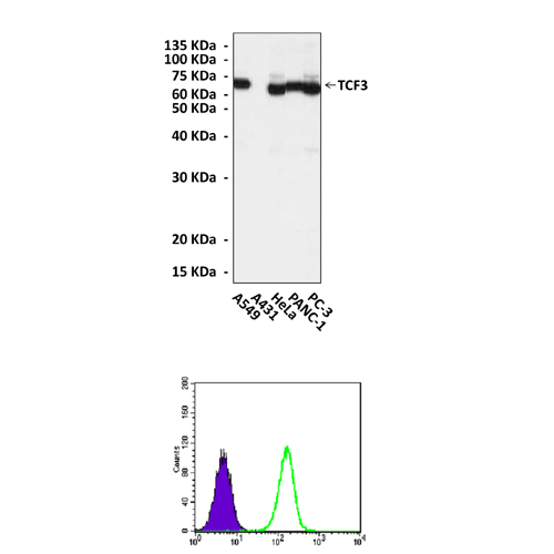
Applications & Suggested starting dilutions:* | WB 1:500 - 1:2000 IP n/d IHC n/d ICC n/d FACS 1:200 - 1:400 |
Predicted Molecular Weight of protein: | 68 kDa |
Specificity/Sensitivity: | Detects endogenous TCF3 proteins without cross-reactivity with other family members. |
Storage: | Store at -20°C, 4°C for frequent use. Avoid repeated freeze-thaw cycles. |
*Optimal working dilutions must be determined by end user.
Products
| Product | Size | CAT.# | Price | Quantity |
|---|---|---|---|---|
| Mouse Tcf3 Antibody: Mouse Tcf3 Antibody | Size: 100 ul | CAT.#: CP10234 | Price: $413.00 |

