Product Sheet CA0360
Description
BACKGROUND Nitric oxide (NO), formed enzymatically from L-arginine, functions as an endogenous signaling molecule in numerous organs and tissues throughout the animal and plant kingdoms. The first NO synthase (NOS) was isolated from mammalian brain and named neuronal NOS (nNOS, aka: NOS1) owing to its localization in neurons. NO plays several important roles in the brain, including in regulation of synaptic signalling and plasticity. Additionally, high levels of nNOS protein are present in skeletal muscle, where NO controls muscle contractility and local blood flow.1 Skeletal muscle nNOS (nNOSm) is a splicing isoform of nNOS that, when translated, results in the addition of a 34 amino acid segment within the reductase domain. nNOS also expressed in cardiac and smooth muscle. nNOS activity is primarily regulated by increases in intracellular Ca2+, which activate nNOS through calmodulin binding. NOS enzymes are homodimeric proteins. Recent studies show that NO actions in brain and muscle also rely crucially upon the association of nNOS with specific protein complexes in neurons and muscle cells, respectively. These physical interactions with nNOS allow for integration of NO signaling into distinct transduction cascades in specific cell types.
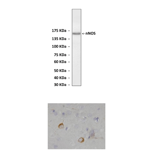
The 160kDa nNOS contains an N-terminal PSD/Discs-large/ZO-1 homologous (PDZ)-binding domain, which anchors this complex to the postsynaptic density in the vicinity of the N-methyl-D-aspartate type-glutamate receptor (NMDAR) in brain. The PDZ domain of nNOS binds to a similar PDZ domain from the postsynaptic density protein, PSD-95, which in turn binds to the cytosolic tail of the NMDAR. These molecular interactions explain how Ca2+ influx through NMDA receptors is efficiently coupled to NO synthesis and activity.2 Following its synthesis at postsynaptic sites, NO may diffuse back to the presynaptic terminal and increase cGMP levels through activation of soluble guanylate cyclase. This membrane-localized nNOS complex is further linked to cytoplasmic signal transduction pathways via the physical interaction of nNOS with DexRas 1 and the adapter protein CAPON, which might activate a downstream MAP kinase cascade and modulate nuclear transcription.3 Functionally, nNOS might also represent a central component that regulates synaptic transmission and intercellular signaling, through negative regulation of the NMDAR by Snitrosylation and NOdependent activation of DexRas. Additionally, the half-life of neuronal nNOS protein is regulated by the Ca2+ sensitive protease calpain. Whereas the small quantities of NO formed during synaptic transmission modulate neuronal signaling, excess NO mediates neurotoxicity in pathological situations, such as an ischemic stroke. This NO toxicity is accentuated in the presence of oxidative radicals such as O2–, which can also be generated by nNOS. Interestingly, nNOS expressing neurons are spared from injury associated with elevated NO, which might partly be because of the physical association of nNOS with phosphofructokinase-M (PFK), the rate-limiting enzyme in glycolysis.4 Consequently, while therapeutic modulation of nNOS represents a potentially important approach in the setting of several clinically important neurological diseases, the balance between positive and negative effects of nNOS derived NO in the brain are complex and must be carefully weighed.
REFERENCES
1. Semenza, G.L.: J. Clin. Invest. 115:2976-8, 2005
2. Cao, J. et al: J. Cell. Biol. 168:117-26, 2005
3. Liu, W. et al: Neurosci. Lett. 410:174-77, 2006
4. Firestein, B.L. & Bredt, D.S.: J. Biol. Chem. 274:10545-50, 1999
2. Cao, J. et al: J. Cell. Biol. 168:117-26, 2005
3. Liu, W. et al: Neurosci. Lett. 410:174-77, 2006
4. Firestein, B.L. & Bredt, D.S.: J. Biol. Chem. 274:10545-50, 1999
Products are for research use only. They are not intended for human, animal, or diagnostic applications.
Details
Cat.No.: | CA0360 |
Antigen: | N-terminal sequence of human NOS1 |
Isotype: | Affinity-purified rabbit polyclonal IgG |
Species & predicted species cross- reactivity ( ): | Human, Rabbit, Rat, Mouse |
Applications & Suggested starting dilutions:* | WB 1:500 - 1:1000 IP n/d IHC (Paraffin) 1:50 - 1:200 ICC n/d FACS n/d |
Predicted Molecular Weight of protein: | 160 kDa |
Specificity/Sensitivity: | Detects endogenous nNOS proteins without cross-reactivity with other family members. |
Storage: | Store at 4° C for frequent use; at -20° C for at least one year. |
*Optimal working dilutions must be determined by end user.
Products
| Product | Size | CAT.# | Price | Quantity |
|---|---|---|---|---|
| Polyclonal Nitric Oxide Synthase 1 (NOS1) Antibody: Polyclonal Nitric Oxide Synthase 1 (NOS1) Antibody | Size: 100 ul | CAT.#: CA0360 | Price: $375.00 |

