Product Sheet CA1343
Description
BACKGROUND Mitogen-activated protein kinases (MAPKs) are serine/threonine kinases that play an instrumental role in signal transduction from the cell surface to the nucleus. MAPKs are major components of pathways controlling embryogenesis, cell differentiation, cell proliferation, and cell death. Mammalian members of this family are extracellular signal-regulated kinases 1/2 (ERK 1/2), c-Jun amino-terminal kinases or stress-activated protein kinases (JNK/SAPKs) and p38 kinases (p38 (MAPK)). MAPKs are regulated by phosphorylation cascades. Two upstream protein kinases activated in series lead to activation of a MAPK, and additional kinases may also be required upstream of this three-kinase module. In all currently known MAPK cascades, the kinase immediately upstream of the MAPK is a member of the MAPK/ERK kinase (MAPKK, MAP2K, MEK or MKK) family. These are dual specificity enzymes that can phosphorylate hydroxyl side chains of serine/threonine and tyrosine residues in their MAPK substrates. In spite of their ability to phosphorylate proteins on both aliphatic and aromatic side chains in the appropriate context, the substrate specificity of the known MEKs is very narrow: each MEK phosphorylates only one or a few of the MAPKs. The MAPK kinases (MEK or MKK) are activated by upstream kinases called MAP kinase kinase kinase (MAPKKK or MAP3K) such as Raf family. The Raf>MEK>MAPK is a well studied signaling pathway up to date.1
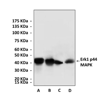
ERK1 and ERK2 are proteins of 44 and 42 kDa that are nearly 85% identical overall, with much greater identity in the core regions involved in binding substrates. ERK1 and ERK2 are activated by a pair of closely related MEKs, MEK1 and MEK2. The two phosphoacceptor sites, tyrosine and threonine, which are phosphorylated to activate the kinases, are separated by a glutamate residue in both ERK1 and ERK2 to give the motif TEY (Thr202/Tyr204 and Thr185/Tyr187 for p44 and p42 respective) in the activation loop. Once activated, Erk1/2 rapidly translocated into the nucleus, where Erk1/2 activates downstream signaling components including Elk-1, although activated Erk1/2 also phosphorylated numerous substrates on (S/T)P sites in all cellular compartments.2 The Erk1/2 was inactivated by dephosphorylation through action of MAPK phosphotase 1 and 2 in nucleus.3
ERK1 and ERK2 are proteins of 44 and 42 kDa that are nearly 85% identical overall, with much greater identity in the core regions involved in binding substrates. ERK1 and ERK2 are activated by a pair of closely related MEKs, MEK1 and MEK2. The two phosphoacceptor sites, tyrosine and threonine, which are phosphorylated to activate the kinases, are separated by a glutamate residue in both ERK1 and ERK2 to give the motif TEY (Thr202/Tyr204 and Thr185/Tyr187 for p44 and p42 respective) in the activation loop. Once activated, Erk1/2 rapidly translocated into the nucleus, where Erk1/2 activates downstream signaling components including Elk-1, although activated Erk1/2 also phosphorylated numerous substrates on (S/T)P sites in all cellular compartments.2 The Erk1/2 was inactivated by dephosphorylation through action of MAPK phosphotase 1 and 2 in nucleus.3
REFERENCES
1. Pearson G et al.: Endocrin. Rev. 22:153-183, 2001.
2. Farooq A & Zhou MM: Cell. Signal. 16:769-779, 2004.
3. Pouyssegur J et al.: Biochem. Pharm. 64:755-763, 2002.
2. Farooq A & Zhou MM: Cell. Signal. 16:769-779, 2004.
3. Pouyssegur J et al.: Biochem. Pharm. 64:755-763, 2002.
Products are for research use only. They are not intended for human, animal, or diagnostic applications.
Details
Cat.No.: | CA1343 |
Antigen: | Short peptide from human Erk1 sequence. |
Isotype: | Rabbit IgG |
Species & predicted species cross- reactivity ( ): | Human, Mouse, Rat |
Applications & Suggested starting dilutions:* | WB 1:1000 IP n/d IHC 1:50 - 1:200 ICC n/d FACS n/d |
Predicted Molecular Weight of protein: | 44 kDa |
Specificity/Sensitivity: | Detects endogenous levels of Erk1 proteins without cross-reactivity with other related proteins. |
Storage: | Store at -20°C, 4°C for frequent use. Avoid repeated freeze-thaw cycles. |
*Optimal working dilutions must be determined by end user.
Products
| Product | Size | CAT.# | Price | Quantity |
|---|---|---|---|---|
| Rabbit Erk1 p44 MAPK Antibody: Rabbit Erk1 p44 MAPK Antibody | Size: 100 ul | CAT.#: CA1343 | Price: $375.00 |

