Product Sheet CP10294
Description
BACKGROUND The Chromobox domain (Cbx) gene family, consisting of Polycomb and Heterochromatin Protein 1 (HP1) genes, is involved in transcriptional repression, cell cycle regulation and chromatin remodeling. The HP1 proteins are transcriptional regulators conserved from Schizosaccharomyces pombe to mammals. These proteins bind histone H3 methylated on lysine 9 (H3K9), an epigenetic mark generated by the histone methyltransferases SU(VAR)3-9 and orthologues, but they also interact with DNA, a wide range of regulatory and structural proteins and a yet uncharacterized RNA component. HP1 proteins are mainly considered to be silencers involved in the spreading of heterochromatin. In mammalian cells, the HP1 family is composed of HP1α (also called CBX5), HP1β (also called CBX1) and HP1γ (also called CBX3). Despite their high degree of sequence homology mutational analysis has revealed different phenotypes indicating that they possess different functions.1 These three isoforms are concentrated in foci of dense pericentromeric heterochromatin but are also present in the rest of the nucleus. Consistent with their general distribution, the mammalian HP1 proteins are detected not only in dense heterochromatic regions but also on active euchromatic genes. For example, HP1β/CBX1 is present on the repressed cyclin E promoter. Similarly, HP1γ/CBX3 participates in the repression of the mouse mammary tumour virus promoter in the absence of hormonal stimulation. Interestingly, HP1γ/CBX3 is also recruited to the coding region of a subset of actively transcribed erythroid-specific genes in a transcription-dependent manner. Moreover, HP1 proteins were also shown to be present on the long terminal repeat (LTR) of HIV1 during the phases of viral latency. This promoter is stimulated by the nuclear factor-κB and the mitogen-activated protein kinase signal-transduction pathways that are both known from other promoters to induce phosphorylation on serine 10 of histone H3 (H3S10). This is noteworthy because phosphorylation of H3S10 is part of the mechanism that causes delocalization of HP1 proteins from the condensing chromosomes during mitosis.2 In addition it was shown that HP1β/CBX1 is involved in DNA damage repair. Minutes after DNA damage, the variant histone H2AX is phosphorylated by protein kinases of the phosphoinositide kinase family, including ATM, ATR or DNA-PK. Phosphorylated (γ)-H2AX, which recruits molecules that sense or signal the presence of DNA breaks, activating the response that leads to repair, is the earliest known marker of chromosomal DNA breakage. DNA breaks swiftly mobilize HP1β/CBX1 bound to histone H3 methylated on lysine 9 (H3K9me). Local changes in histone-tail modifications are not apparent. Instead, phosphorylation of HP1β/CBX1 on amino acid Thr 51 accompanies mobilization, releasing HP1β/CBX1 from chromatin by disrupting hydrogen bonds that fold its chromodomain around H3K9me. Inhibition of casein kinase 2 (CK2), an enzyme implicated in DNA damage sensing and repair, suppresses Thr 51 phosphorylation and HP1β/CBX1 mobilization in living cells. CK2 inhibition, or a constitutively chromatin-bound HP1β/CBX1 mutant, diminishes H2AX phosphorylation.3 Moreover, other studies showed that N-terminal phosphorylation of HP1α/CBX5 promotes its chromatin binding.4 Thus, HP1 signaling cascade helps to initiate the biological response, altering chromatin by modifying a histone-code mediator protein, but not the code itself. Loss of HP1 proteins causes chromosome segregation defects and lethality in some organisms; a reduction in levels of HP1 family members is associated with cancer progression in humans. These consequences are likely due to the role of HP1 in centromere stability, telomere capping and the regulation of euchromatic and heterochromatic gene expression.5
REFERENCES
1. Singh, P.B.: Russia J. Genet. 46:1257-62, 2010
2. Mateescu, B. et al: EMBO Report 9:267-72, 2008
3. Ayoub, N. et al: Nature 453:682-6, 2008
4. Hiragami-Hamada, K. et al: Mol. Cell. Biol. 2011 (in press)
5. Billur, M. et al: Trends Biol. Chem. 35:115-23, 2010
2. Mateescu, B. et al: EMBO Report 9:267-72, 2008
3. Ayoub, N. et al: Nature 453:682-6, 2008
4. Hiragami-Hamada, K. et al: Mol. Cell. Biol. 2011 (in press)
5. Billur, M. et al: Trends Biol. Chem. 35:115-23, 2010
Products are for research use only. They are not intended for human, animal, or diagnostic applications.

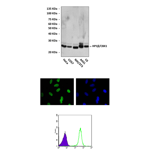
(Click to Enlarge) Top: Western Blot detection of HP1beta/CBX1 proteins in various cell lysates using HP1beta/CBX1 Antibody. Middle: This antibody stains HeLa cells in immunofluorescent analysis (Cyclin B1 Antibody: Green; DRAQ5 DNA Dye: Blue). Bottom: It also specifically reacts with HP1beta/CBX1 proteins in Cos7 cells by FACS testing (HP1beta/CBX1 Antibody: Green; control; Purple).
Details
Cat.No.: | CP10294 |
Antigen: | Raised against recombinant human HP1β/CBX1 fragments expressed in E. coli. |
Isotype: | Mouse IgG1 |
Species & predicted species cross- reactivity ( ): | Human, Mouse, Rat |
Applications & Suggested starting dilutions:* | WB 1:1000 IP n/d IHC n/d ICC 1:50 - 1:200 FACS 1:50 - 1:200 |
Predicted Molecular Weight of protein: | 26 kDa |
Specificity/Sensitivity: | Detects endogenous HP1β/CBX1 proteins without cross-reactivity with other family members. |
Storage: | Store at -20°C, 4°C for frequent use. Avoid repeated freeze-thaw cycles. |
*Optimal working dilutions must be determined by end user.
Products
| Product | Size | CAT.# | Price | Quantity |
|---|---|---|---|---|
| Mouse HP1Beta/CBX1 Antibody: Mouse HP1beta/CBX1 Antibody | Size: 100 ul | CAT.#: CP10294 | Price: $413.00 |
