Product Sheet CP10310
Description
BACKGROUND Methionine adenosyltransferase (MAT, ATP:L-methionine Sadenosyltransferase, EC 2.5.1.6) catalyzes the biosynthesis of S-adenosylmethionine (AdoMet) from methionine and ATP and thus plays a central role in cellular metabolism. The the adenosyl moiety of ATP is transferred to methionine, forming a highly energetic compound as a result of a sulphonium bond between the 5’-carbon atom of the ribose and the sulphur atom of the amino acid. There are two isoenzymes of MAT: one is liver-specific and the other is present in all tissues. The first has a relatively high Km for methionine and the second has a low Km for methionine.1 AdoMet acts as the methyl donor for over 115 different cellular transmethylation reactions, including those of DNA, RNA, proteins, and lipids. DNA methylation is a important epigenetic feature of DNA that plays a critical role in gene expression and regulation. In addition, AdoMet participates in the transsulfuration pathway and, after decarboxylation, serves as a propylamine group donor in the biosynthesis of polyamines.2 Moreover, with the transfer of methyl group, AdoMet is converted into S-adenosylhomocysteine (AdoHcy). The majority of AdoMet-dependent methyltransferases are strongly inhibited by AdoHcy. The intracellular AdoMet/AdoHcy ratio has been used as a predictor of cellular methylation activity. AdoHcy is further converted into homocysteine and adenosine by the AdoHcy hydrolase, which is widely distributed in mammalian tissues. Hypercysteinaemia has been regarded as a new modifiable risk factor for atherosclerosis and vascular diseases.3
REFERENCES
1. Castro, R. et al: J. Inherit. Metb. Dis. 29:3-20, 2006
2. Ubagai, T. et al: J. Clin. Invest. 96:1943-7, 1995
3. Fowler, B.: Semin. .Vascul. Med. 5:77-86, 2005
2. Ubagai, T. et al: J. Clin. Invest. 96:1943-7, 1995
3. Fowler, B.: Semin. .Vascul. Med. 5:77-86, 2005
Products are for research use only. They are not intended for human, animal, or diagnostic applications.

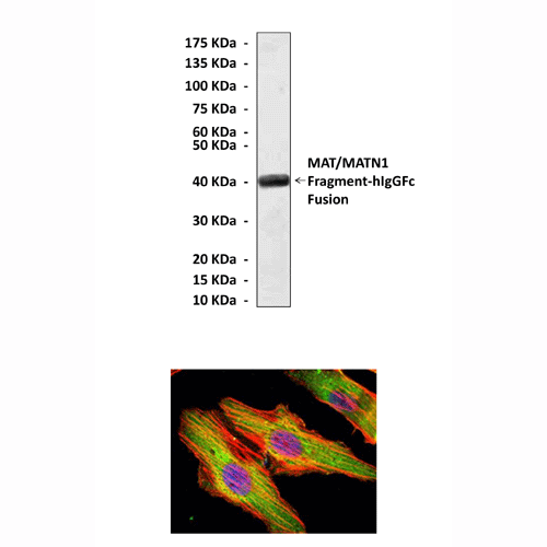
(Click to Enlarge) Top: Western Blot detection of MAT/MATN1 proteins in cell lysates from 293 cells and 293 cells transfected with human MAT fragment-hIgGFc fusion-expressing vectors using MAT/MATN1 Antibody. Bottom: This antibody stains HeLa cells in confocal immunofluorescent analysis (MAT/MATN1 Antibody: Green; Actin filaments: Red; DRAQ5 DNA Dye: Blue).
Details
Cat.No.: | CP10310 |
Antigen: | Raised against recombinant human MAT/MATN1 fragments expressed in E. coli. |
Isotype: | Mouse IgG1 |
Species & predicted species cross- reactivity ( ): | Human, Mouse, Rat |
Applications & Suggested starting dilutions:* | WB 1:1000 IP 1:50 IHC n/d ICC 1:50 - 1:200 FACS n/d |
Predicted Molecular Weight of protein: | 54 kDa |
Specificity/Sensitivity: | Detects endogenous MAT/MATN1 proteins without cross-reactivity with other family members. |
Storage: | Store at -20°C, 4°C for frequent use. Avoid repeated freeze-thaw cycles. |
*Optimal working dilutions must be determined by end user.a
Products
| Product | Size | CAT.# | Price | Quantity |
|---|---|---|---|---|
| Mouse MAT/MATN1 Antibody: Mouse MAT/MATN1 Antibody | Size: 100 ul | CAT.#: CP10310 | Price: $413.00 |
