Product Sheet CP10410
Description
BACKGROUND Prohibitins belong to the Band-7 family of proteins and are ubiquitously expressed, multifunctional proteins implicated in cellular processes including mitochondrial function and protein folding, proliferation control and suppression of oncogenesis, apoptosis, and transcription regulation. They are mainly localize to mitochondria. The eukaryotic mitochondrial prohibitin (PHB) complex comprises two highly homologous subunits, PHB-1 and PHB-2 (around 50% amino acid sequence identity and 60% similarity). PHB-1 and PHB-2, with molecular weights of 32 and 34 kDa respectively, associate to form a ring-like macromolecular structure of approximately 1 MD with a diameter of 20-25 nm. This high molecular weight complex has been identified inyeast, Caenorhabditis elegans and mammals. PHB contains an N-terminal hydrophobic membrane anchoring α1-helix domain and a C-terminal leucine/isoleucine-rich motif that acts as a nuclear export signal. PHB-1 and PHB-2 are interdependent for protein complex formation, and elimination of either PHB-1 or PHB-2 results in the absence of the whole PHB complex. The PHB complex sits at the mitochondrial inner membrane facing the inter membrane space. Several roles have been proposed for mitochondrial prohibitins. First, the PHB complex was suggested to regulate membrane protein degradation by the mitochondrial m-AAA protease. Later, a function as a membrane-bound chaperone, which holds and stabilizes newly synthesized mitochondrial-encoded proteins was proposed. PHB proteins might also play a role in stabilizing the mitochondrial genome. In addition, the PHB complex has been implicated in mitochondrial morphogenesis by stabilizing OPA-1, and by functioning as scaffold proteins that recruit membrane proteins to a specific lipid environment.1 In addition, PHB-1 also presents in the nucleus, involved in regulation of transcription. Moreover, it can be found in the plasma membrane and cytoplasm. It has been demonstrated that PHB-1 is involved in phophatidylinositol-3-kinase (PI3K)/protein kinase B (Akt) and transforming growth factor-beta (TGF-beta)/signal transducers and activators of transcription signaling pathways, and Ras/mitogen-activated protein kinase (MAPK)/extracellular signal-regulated kinase (ERK) signaling. PHB-1 might also function as a cell-surface receptor for an as-yet unidentified ligand. Cell-associated PHB-1 in the gastrointestinal tract has been implicated in protection against infection and inflammation and the induction of apoptosis in other tissues. In addition, Acting as a binding site for ubiquitin, prohibitin protein regulates protein degradation by proteasome. Moreover, it was shown that prohibitin recruits chromatin-remodeling molecules to gene promoter elements for transcriptional repression. In addition to transcriptional repression, prohibitin can induce p53-mediated transcription, indicating that prohibitin may have dual functions in modulating transcription.2 In recent years, over-expression of prohibitin has been detected in some tumor cells, including lung cancer, prostate cancer, cervical cancer, bladder cancer, gastric cancer, and breast cancer. Moreover, prohibitin may exert different functional roles, it has a permissive action on tumor growth or acts as an oncosuppressor. The diverse array of functions of PHB-1, together with the emerging evidence that its function can be modulated specifically in certain tissue, suggest that targeting PHB-1 would be a useful therapeutic approach for the treatment of variety of disease states, including inflammation, obesity and cancer.3
REFERENCES
1. Wang, S. & Faller, D.V.: Transl. Oncogenomics 3:23–7, 2008
2. Theiss, A.L. et al: Mol. Biol. Cell. 20:4412–23, 2009
3. Jia, L. et al: Neoplasma 58:104-9, 2011
2. Theiss, A.L. et al: Mol. Biol. Cell. 20:4412–23, 2009
3. Jia, L. et al: Neoplasma 58:104-9, 2011
Products are for research use only. They are not intended for human, animal, or diagnostic applications.

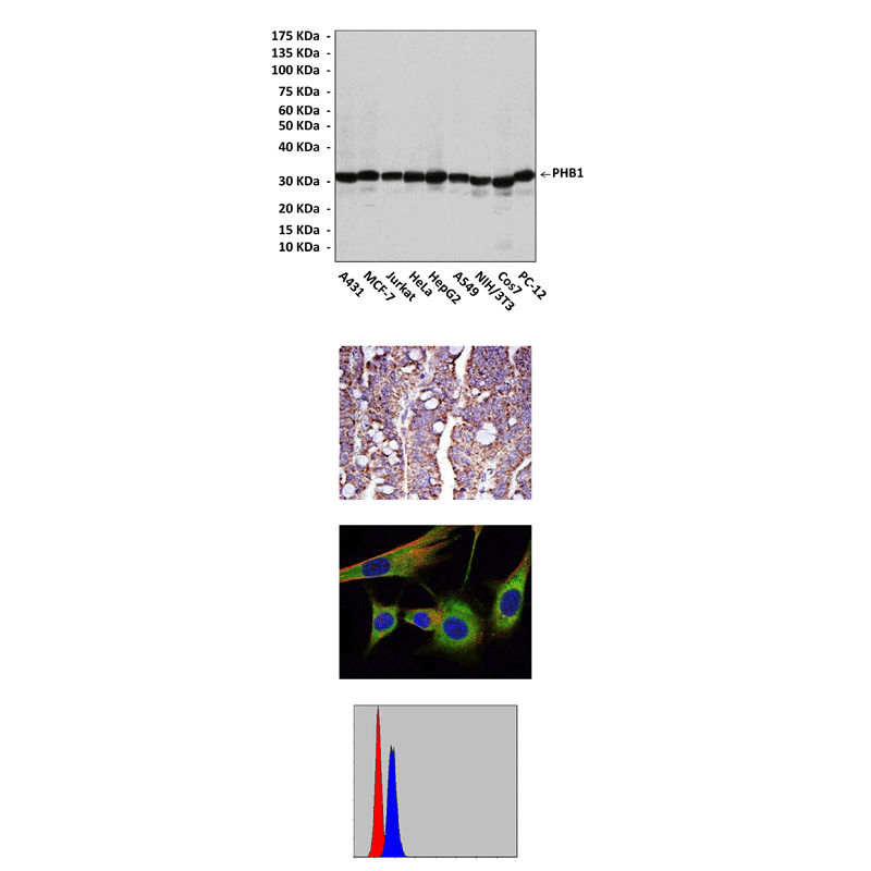
(Click to Enlarge) Top: Western blot detection of PHB1 proteins in various cell lysates using PHB1 Antibody. Middle, Upper: It also stains paraffin-embedded human rectum cancer tissue in IHC analysis. Middle, Lower: This antibody stains NIH3T3 cells in confocal immunofluorescent testing (PHB1 Antibody: Green; Actin filaments: Red; DRAQ5 DNA Dye: Blue). Bottom: This antibody detects PHB1 proteins specifically in MCF7 cells by FACS assay (PHB1 Antibody: Blue; negative control: Red).
Details
Cat.No.: | CP10410 |
Antigen: | Raised against recombinant human PHB-1 fragments expressed in E. coli. |
Isotype: | Mouse IgG1 |
Species & predicted species cross- reactivity ( ): | Human, Mouse, Rat |
Applications & Suggested starting dilutions:* | WB 1:1000 IP n/d IHC 1:50 - 1:200 ICC 1:50 - 1:200 FACS 1:50 - 1:200 |
Predicted Molecular Weight of protein: | 32 kDa |
Specificity/Sensitivity: | Detects PHB-1 proteins in various cell lysate. |
Storage: | Store at -20°C, 4°C for frequent use. Avoid repeated freeze-thaw cycles. |
*Optimal working dilutions must be determined by end user.
Products
| Product | Size | CAT.# | Price | Quantity |
|---|---|---|---|---|
| Mouse PHB1 Antibody: Mouse PHB1 Antibody | Size: 100 ul | CAT.#: CP10410 | Price: $457.00 |
