Product Sheet CC1033
Description
BACKGROUND RET (Rearranged during Transfection) encodes a receptor tyrosine kinase with key roles in cell growth, differentiation, and survival. It consists of three functional domains, an extracellular domain with four cadherin-like repeats, a cysteine-rich region, and an intracellular tyrosine kinase region . Following ligand binding to the extracellular region, receptor dimerization is mediated by activation of the cysteine-rich region that lies adjacent to the plasma membrane. Receptor dimerization leads to autophosphorylation of intracellular tyrosine residues, which subsequently activate downstream pathways of signal transduction.1 Ligands for RET include those in the glial cell-line derived neurotrophic factor (GDNF) family, including persepherin, artemin, and neurturin. RET signals through multiple downstream pathways. One such pathway, the RAS/MEK/ERK pathway promotes cell cycle progression; another downstream pathway, P13K/AKT/NF-κB, leads to increased cell motility, survival, and progression through the cell cycle.2 RET activation also stimulates p38, MAPK, JAK/STAT, and protein kinase C.3 RET is expressed widely in neural-crest derived tissues such as noradrenergic and dopamanergic neurons and neuroendocrine tissues including thyroid C cells and the adrenal medulla. RET is also involved in the development of the enteric nervous system and the kidney.
Activating RET germline mutations have been identified as the primary cause of the hereditary medullary thyroid carcinoma.3 The RET proto-oncogene has become the rational target for molecularly designed drug therapy.
REFERENCES
1. Mak, Y.F. & Ponder, B.A.J.: Curr. Opin. Genet. Develop. 6:82-86, 1996.
2. Arighi, E. et al.: Cytokine Growth Factor Rev. 16: 441–67, 2005.
3. Santoro, M. et al.: Eur. J Endocrinol. 133: 513-522, 1995.
2. Arighi, E. et al.: Cytokine Growth Factor Rev. 16: 441–67, 2005.
3. Santoro, M. et al.: Eur. J Endocrinol. 133: 513-522, 1995.
Products are for research use only. They are not intended for human, animal, or diagnostic applications.
Details
Cat.No.: | CC1033 |
Antigen: | sequence near the human Ret carboxyl terminal. |
Isotype: | Affinity purified rabbit IgG |
Species & predicted species cross- reactivity ( ): | Human |
Applications & Suggested starting dilutions: | WB 1:1000 IP n/d IHC (Paraffin) n/d ICC n/d FACS n/d |
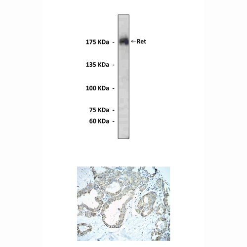
Predicted Molecular Weight of protein: | 175 kDa |
Specificity/Sensitivity: | This antibody detects human Ret proteins in transfected cell lysate. |
Storage: | Store at 4° C for frequent use; at -20° C for at least one year. |
*Optimal working dilutions must be determined by end user.
Products
| Product | Size | CAT.# | Price | Quantity |
|---|---|---|---|---|
| Polyclonal Ret Antibody: Polyclonal Ret Antibody | Size: 100 ul | CAT.#: CC1033 | Price: $333.00 |

