Product Sheet CG1127
Description
BACKGROUND The c-erbB family of receptors includes four distinct receptors, namely c-erb B1 (EGF receptor), 2, 3 and 4 (HER1, 2, 3 and 4, respectively). The epidermal growth factor (EGF) and related peptides bind the ErbB receptors, inducing the formation of different homo- and heterodimers. Receptor dimerization promotes activation of the intrinsic kinase, leading to phosphorylation of specific tyrosines located in the ErbB\'s cytoplasmic region. These phosphorylated residues serve as docking sites for a variety of signaling molecules whose recruitment stimulates intracellular signaling cascades, which ultimately control diverse genetic programs.1 In contrast to other receptor tyrosine kinases; the kinase domain of EGFR (ERBB1) does not require phosphorylation for activation. Consequently, the overall activation state of the receptor is controlled by constant balancing of activity favoring and activity suppressing actions within the receptor molecule. Influences of the membrane microenvironment and context dependent interactions with varying sets of signaling partners are superimposed on this system of intramolecular checks and balances.2
It was shown that EGFR overexpression and mutations occurred in many tumors. The EGFR is known to be involved in carcinogenetic processes such as cell proliferation, apoptosis, angiogenesis, cell motility, and metastasis. Preclinical and clinical studies have shown that targeting EGFR is a valid method for anticancer therapy.3 Strategies aimed at inhibiting the EGFR pathway included different classes of compounds, with monoclonal antibodies and tyrosine kinase inhibitors being the most widely-investigated agents in cancer therapy.
It was shown that EGFR overexpression and mutations occurred in many tumors. The EGFR is known to be involved in carcinogenetic processes such as cell proliferation, apoptosis, angiogenesis, cell motility, and metastasis. Preclinical and clinical studies have shown that targeting EGFR is a valid method for anticancer therapy.3 Strategies aimed at inhibiting the EGFR pathway included different classes of compounds, with monoclonal antibodies and tyrosine kinase inhibitors being the most widely-investigated agents in cancer therapy.
REFERENCES
1. Holbro, T. & Hynes, N. E.: Annu Rev Pharmacol Toxicol. 44:195, 2004.
2. Warren, C.M. & Landgraf, R.: Cell Signal 18:923, 2006.
3. Arteaga, C. L.: Semin Oncol. 29(5 Suppl 14):3, 2002.
2. Warren, C.M. & Landgraf, R.: Cell Signal 18:923, 2006.
3. Arteaga, C. L.: Semin Oncol. 29(5 Suppl 14):3, 2002.
Products are for research use only. They are not intended for human, animal, or diagnostic applications.

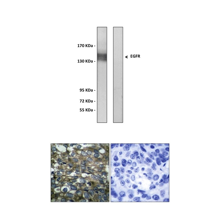
(Click to Enlarge) Top: Immunoblotting analysis of extracts from A431 cells, treated with EGF 40?M 10′, using Anti-EGFR antibody. The lane on the left was treated with the Anti-EGFR antibody. The lane on the right (negative control) was treated with both Anti-EGFR antibody and the synthesized immunogen peptide. Bottom: Immunohistochemistry analysis of paraffin-embedded human breast carcinoma tissue using Anti-EGFR antibody. Cells on the left were treated with the Anti-EGFR antibody. Cells on the right (negative control) were treated with both Anti-EGFR antibody and the synthesized immunogen peptide.
Details
Cat.No.: | CG1127 |
Antigen: | Synthesized peptide derived from human EGFR. |
Isotype: | Rabbit IgG |
Species & predicted species cross- reactivity ( ): | Human, Mouse, Rat |
Applications & Suggested starting dilutions:* | WB 1:500-1:1000 IP n/d IHC 1:50-:100 ICC n/d FACS n/d |
Predicted Molecular Weight of protein: | 134 kDa |
Specificity/Sensitivity: | Detects endogenous EGFR proteins without cross-reactivity with other family members. |
Storage: | Store at -20°C, 4°C for frequent use. Avoid repeated freeze-thaw cycles. |
*Optimal working dilutions must be determined by end user.
Products
| Product | Size | CAT.# | Price | Quantity |
|---|---|---|---|---|
| Rabbit EGFR Antibody: Rabbit EGFR Antibody | Size: 100 ul | CAT.#: CG1127 | Price: $384.00 |
