Product Sheet CP10099
Description
BACKGROUND The Fujinami poultry sarcoma/feline sarcoma (fps/fes) proto-oncogene encodes a structurally unique nonreceptor protein tyrosine kinase (PTK) called Fps/Fes kinase. FER kinase (94 kDa) is the only other known member of this distinct subfamily in mammalian cells. In contrast to the tissue-specific expression pattern of Fps/Fes kinase, FER kinase is a ubiquitously expressed nonreceptor PTK-associated with cell-cell actin-based adherens junction (AJ) - and intermediate filament-basedl desmosomes. Fps/Fes and FER kinases have been implicated in the regulation of cell-cell AJs and cell-matrix focal adhesion interactions in multiple epithelia, and they serve similar and even redundant biological function based on recently completed genetic analysis in mice.1 There is a truncated FER, FerT kinase (51 kDa) in testis. The FCH (Fps/Fes/FER/ClP4 homology) and the three coiled-coil (CC) domains from the N-terminus of FER kinase are missing from FerT but FerT has a unique N-terminal sequence of 44 amino acid residues.2
FER kinase is a cytoplasmic PTK found at the site of AJs and was shown to associate with the phosphorylated form of p120ctn, which in turn interacts with the cadherin/catenin complex. As such, FER kinase may be an important PTK that mediates signals at the site of AJs to the nucleus in much the same way as p120ctn and beta-catenin. FER kinase is an important regulator of cell adhesion function. For instance, overexpression of FER kinase in embryonic fibroblasts can reduce the level of alpha-catenin and beta-catenin associating with E-cadherin, causing a loss of cell adhesion function. Other studies have shown that FER kinase can induce phosphorylation of p120ctn and beta-catenin, dissociating them from the cadherin/catenin complex, causing a loss of cell adhesion function. Furthermore, FER kinase mediates cross-talk between cadherin and beta1-integrin.3
REFERENCES
1. Greer, P. et al: Nature Rev. Mol. Cell. Biol. 3:278-89, 2002
2. Hazan, B. et al: Cell. Grow. Diff.4:443-9, 1993
3. Chen, Y-M. et al: Biol. Reproduct. 69:656-72, 2003
2. Hazan, B. et al: Cell. Grow. Diff.4:443-9, 1993
3. Chen, Y-M. et al: Biol. Reproduct. 69:656-72, 2003
Products are for research use only. They are not intended for human, animal, or diagnostic applications.

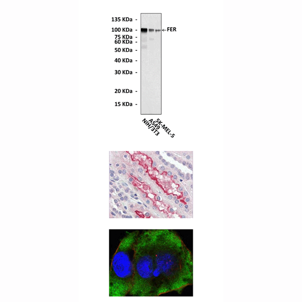
(Click to Enlarge) Top: Western Blot detection of FER proteins in various cell lysates using FER Antibody. Middle: This antibody stains paraffin-embedded human kidney tissue in immunohistochemical analysis. Bottom: It also stains HeLa cells in confocal immunofluorescent testing (FER Antibody: Green; Actin filament: Red, DAQR5 DNA dye: Blue).
Details
Cat.No.: | CP10099 |
Antigen: | Purified recombinant human FER fragments expressed in E. coli. |
Isotype: | Mouse IgG |
Species & predicted species cross- reactivity ( ): | Human, Mouse, Rat |
Applications & Suggested starting dilutions:* | WB 1:1000 IP n/d IHC 1:200 ICC 1:200 FACS n/d |
Predicted Molecular Weight of protein: | 94 kDa |
Specificity/Sensitivity: | Detects endogenous FER proteins without cross-reactivity with other family members. |
Storage: | Store at -20°C, 4°C for frequent use. Avoid repeated freeze-thaw cycles. |
*Optimal working dilutions must be determined by end user.
Products
| Product | Size | CAT.# | Price | Quantity |
|---|---|---|---|---|
| Mouse Fer Tyrosine Kinase Antibody: Mouse Fer Tyrosine Kinase Antibody | Size: 100 ul | CAT.#: CP10099 | Price: $472.00 |
