Product Sheet CP10167
Description
BACKGROUND Chemokines (Chemotactic Cytokines) are small heparin-binding proteins, which constitute a large family of peptides (60-100 amino acids) structurally related to cytokines whose main function is to regulate cell trafficking. They are subdivided into four families based on the number and spacing of the conserved cysteine residues in the N-terminus of the protein: CXC, CC, CX3C, and C, in agreement with the systematic nomenclature. Chemokines play a major role in selectively recruiting monocytes, neutrophils and lymphocytes as well as inducing chemotaxis through the activation of G-protein-coupled receptors (GPCR).1
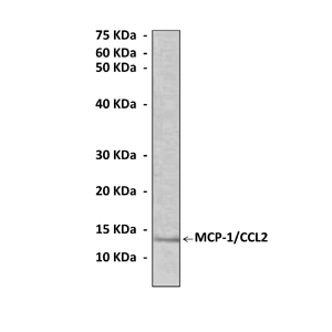
Monocyte Chemoattractant Protein-1 (MCP-1) was the first CC Chemokine identified. Human MCP-1 is composed of 76 amino acids and is 13 kDa in size. MCP subfamily composed of at least four members (MCP-1, -2, -3, and -4).MCP-1 is produced by a variety of cell types, either constitutively or after induction by oxidative stress, cytokines, or growth factors. Many activities have been assigned to MCP-1, including induction of migration of macrophages/monocytes, memory/activated T cells and NK cells and activation of mast cells. MCP-1 mediates its cellular effects primarily through its binding to CCR2, which exists in A and B forms that arise via alternative splicing of the carboxyl-terminal tail. Unlike MCP-1, CCR2 expression is relatively restricted to certain types of cells. CCR2A is the major isoform expressed by mononuclear cells and vascular smooth muscle cells, while monocytes and activated NK cells, express predominantly the CCR2B isoform. It is possible that CCR2A and CCR2B may activate different signaling pathway and exert different actions. In addition, there may be another alternative MCP-1 receptor which may be important in mediating some of the effects of MCP-1 in atherosclerotic arteries and in other inflammatory processes.2 Both MCP-1 and its receptor CCR2 have been demonstrated to be induced and involved in a variety of diseases. Migration of monocytes from the blood stream across the vascular endothelium is required for routine immunological surveillance of tissues, as well as in response to inflammation.
Analysis of the signal transduction pathways triggered by MCP-1 has revealed that it induces a pertussis toxin (PTX)-sensitive rise of intracellular calcium, inhibition of adenyl cyclase, phospholipase C activation, activation of extracellular signal-related kinases (ERKs) and the 2 stress-activated protein kinases (SAPKs), JNK1 and P38, stimulation of 2 separate PI 3-kinase isoforms, namely p85/p110 PI3-kinase (PI3-K) and PI3K-C2 . Recently, analysis of signaling events in human monocytic cells has shown that MCP-1 triggers tyrosine phosphorylation and activation of the JAK2/STAT3 pathway in a PTX-independent manner. Importantly, the signaling pathways through which MCP-1 triggers cell migration are still poorly documented. The exact relationships between ERKs, PI3-K, and transient calcium influx in inducing leukocyte migration in response to MCP-1 remains elusive.3 THP-1 cells were stimulated with MCP-1 and a strong activation of PI 3-kinase at the leading edge was observed. This is consistent with general requirement of PI 3-kinase activation in cell migration.4 The localized accumulation of PIP3 defines the migration direction and triggers the activation of the Rho-family GTPase at the leading edge. Rho-family GTPases are critical molecular switches that modulate the actin cytoskeleton. For example, RhoA triggers actin stress fiber formation; Rac1 induces lamellipodia and membrane ruffling; and Cdc42 elicits the formation of filopodia, i.e., protrusive actin.5
REFERENCES
1. Gerard, C. & and Rollins, B.J.: Nature Immunol. 2:108-115, 2001
2. Schecter, S.D. et al: J. Leuk. Biol. 75:1079-1085, 2004
3. Cambien, B. et al: Blood 97:359-66, 2001
4. Volpe, S. et al: PLoS ONE 5: e10159, 2010
5. Du, D. et al: Dev. Cell 18:52-63, 2010.
2. Schecter, S.D. et al: J. Leuk. Biol. 75:1079-1085, 2004
3. Cambien, B. et al: Blood 97:359-66, 2001
4. Volpe, S. et al: PLoS ONE 5: e10159, 2010
5. Du, D. et al: Dev. Cell 18:52-63, 2010.
Products are for research use only. They are not intended for human, animal, or diagnostic applications.
Details
Cat.No.: | CP10167 |
Antigen: | Purified recombinant human MCP-1 proteins expressed in E. coli. |
Isotype: | Mouse IgG1 |
Species & predicted species cross- reactivity ( ): | Human, Mouse, Rat |
Applications & Suggested starting dilutions:* | WB 1:1000 IP 1:50 IHC n/d ICC n/d FACS n/d |
Predicted Molecular Weight of protein: | 13 kDa |
Specificity/Sensitivity: | Detects MCP-1 proteins without cross-reactivity with other family members. |
Storage: | Store at -20°C, 4°C for frequent use. Avoid repeated freeze-thaw cycles. |
*Optimal working dilutions must be determined by end user.
Products
| Product | Size | CAT.# | Price | Quantity |
|---|---|---|---|---|
| Mouse MCP-1/CCL2 Antibody: Mouse MCP-1/CCL2 Antibody | Size: 100 ul | CAT.#: CP10167 | Price: $354.00 |

