Product Sheet CP10252
Description
BACKGROUND Abl tyrosine kinase family has two members: Abl1 (also named c-Abl) and Abl2 ( also named ARG). Each Abl protein contains an SH3-SH2-TK (Src homology 3-Src homology 2-tyrosine kinase) domain cassette, which confers autoregulated kinase activity and is common among nonreceptor tyrosine kinases. This cassette is coupled to an actin-binding and -bundling domain, which makes Abl proteins capable of connecting phosphoregulation with actin-filament reorganization. Both of these genes are protooncogene and have a highly identity in their structures especially in SH2, SH3, N terminal and tyrosine kinase domain, but the C terminal of these molecules has less than 30% identity. Both Abl1 and Abl2 have evolved to perform specialized functions. Abl1 includes nuclear localization signals and a DNA binding domain through which it mediates DNA damage-repair functions, whereas Abl2 has additional binding capacity for actin and for microtubules to enhance its cytoskeletal remodeling functions. Several types of posttranslational modifications control Abl catalytic activity, subcellular localization, and stability, with consequences for both cytoplasmic and nuclear Abl functions. Binding partners provide additional regulation of Abl catalytic activity, substrate specificity, and downstream signaling. Information on Abl regulatory mechanisms is being mined to provide new therapeutic strategies against hematopoietic malignancies caused by BCR-ABL1 and related leukemogenic proteins.1
Abl2 is involved in homologous recombination, DNA repair or cell cycle progression mechanisms. This protein can interact with c-abl and response to oxidative stress. Like c-abl, Abl2 seems to have a role in apoptosis via phosphorilation of the apoptotic Siva1 protein, control of cell motility and morphogenesis by modifying F-actin structure. Both proteins are also required for maintenance of cortical dendreties in brain. Abl2 is important for development of nervous system and for brain function in adult tissue. Mice with disruption of Abl2 can develop normally but display behavioral disorders. Through its interactions with the cytoskeleton Abl2 plays a role in murine neurulation and it is required for adhesion-dependent neurite branching and synapse/dendrite stability. Abl2 has also a role in fibroblastic- and epithelial cell adhesion and migration. Abl2 shares a number of cellular regulatory functions with c-Abl. Targeted disruption of the c-Abl gene in mice resulted in pleiotropic phenotypes including runtedness, high perinatal lethality, susceptibility to infections, and immune deficiencies. In contrast, homozygous Abl2 knockout mice are healthy and exhibit no immune-deficient phenotypes. Moreover, it was demonstrated that Abl2 and c-abl are differently expressed in B lymphoid cells at different stages of differentiation.2 Unlike Abl2, the level of c-abl transcripts was not related to differentiation. The highest level of Abl2 was found in mature B lymphoid cells (Raji cell line) in contrast to early pre B lymphoid cells. The increment of Abl2 level was also reported in other studies on granulocytes, monocytes and neuron. These findings support the hypothesis that high level of Abl2 is associated to the mature state of specific cells. However, evidence for overlapping roles of c-Abl and Abl2 was obtained by analyzing mice deficient for both genes, which revealed that both proteins are required for normal mouse development. Abl/Abl2-/- mice die around embryonic day 10.5, and the embryos display bleeding in the pericardial sac, exhibit reduced proliferation and cytokine production in response to TCR stimulation and showed a significantly impaired antibody production. Like c-Abl, Abl2 is involved in human neoplastic diseases. Abl is up- or down-regulated in several solid tumors and oncogenic gene translocations involving the ETV6 gene have been described in human acute leukemia.3
Abl2 is involved in homologous recombination, DNA repair or cell cycle progression mechanisms. This protein can interact with c-abl and response to oxidative stress. Like c-abl, Abl2 seems to have a role in apoptosis via phosphorilation of the apoptotic Siva1 protein, control of cell motility and morphogenesis by modifying F-actin structure. Both proteins are also required for maintenance of cortical dendreties in brain. Abl2 is important for development of nervous system and for brain function in adult tissue. Mice with disruption of Abl2 can develop normally but display behavioral disorders. Through its interactions with the cytoskeleton Abl2 plays a role in murine neurulation and it is required for adhesion-dependent neurite branching and synapse/dendrite stability. Abl2 has also a role in fibroblastic- and epithelial cell adhesion and migration. Abl2 shares a number of cellular regulatory functions with c-Abl. Targeted disruption of the c-Abl gene in mice resulted in pleiotropic phenotypes including runtedness, high perinatal lethality, susceptibility to infections, and immune deficiencies. In contrast, homozygous Abl2 knockout mice are healthy and exhibit no immune-deficient phenotypes. Moreover, it was demonstrated that Abl2 and c-abl are differently expressed in B lymphoid cells at different stages of differentiation.2 Unlike Abl2, the level of c-abl transcripts was not related to differentiation. The highest level of Abl2 was found in mature B lymphoid cells (Raji cell line) in contrast to early pre B lymphoid cells. The increment of Abl2 level was also reported in other studies on granulocytes, monocytes and neuron. These findings support the hypothesis that high level of Abl2 is associated to the mature state of specific cells. However, evidence for overlapping roles of c-Abl and Abl2 was obtained by analyzing mice deficient for both genes, which revealed that both proteins are required for normal mouse development. Abl/Abl2-/- mice die around embryonic day 10.5, and the embryos display bleeding in the pericardial sac, exhibit reduced proliferation and cytokine production in response to TCR stimulation and showed a significantly impaired antibody production. Like c-Abl, Abl2 is involved in human neoplastic diseases. Abl is up- or down-regulated in several solid tumors and oncogenic gene translocations involving the ETV6 gene have been described in human acute leukemia.3
REFERENCES
1. Colicelli, J.: Sci. Signal 3:re3, 2010
2. Shimizu, A. et al: J. Biol. Chem. 283:27230-8, 2008
3. Iijima, Y. et al: Blood 95: 2126-31, 2000
2. Shimizu, A. et al: J. Biol. Chem. 283:27230-8, 2008
3. Iijima, Y. et al: Blood 95: 2126-31, 2000
Products are for research use only. They are not intended for human, animal, or diagnostic applications.
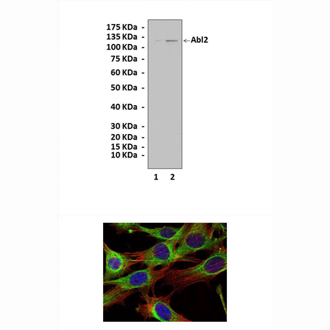
(Click to Enlarge) Top: Western Blot detection of Abl2 proteins in cell lysates from 293 cell transfected with mock vector (lane 1) or human Abl2-expression vector (lane 2) using Abl2 Antibody. Bottom: This antibody also stains NIH3T3 cells in confocal immunofluorescent analysis (Abl2 antibody: Green; Actin filaments: Red; and DRAQ5: blue).
Details
Cat.No.: | CP10252 |
Antigen: | Purified recombinant human Abl2 fragments expressed in E. coli. |
Isotype: | Mouse IgG1 |
Species & predicted species cross- reactivity ( ): | Human, Mouse, Rat |
Applications & Suggested starting dilutions:* | WB 1:1000 IP 1:50 IHC n/d ICC 1:50 - 1:200 FACS n/d |
Predicted Molecular Weight of protein: | 128 kDa |
Specificity/Sensitivity: | Detects Abl2 proteins without cross-reactivity with other related proteins. |
Storage: | Store at -20°C, 4°C for frequent use. Avoid repeated freeze-thaw cycles. |
*Optimal working dilutions must be determined by end user.
Products
| Product | Size | CAT.# | Price | Quantity |
|---|---|---|---|---|
| Mouse Abl2 Antibody: Mouse Abl2 Antibody | Size: 100 ul | CAT.#: CP10252 | Price: $413.00 |