Product Sheet CP10400
Description
BACKGROUND Numerous pro-apoptotic signal transducing molecules act on mitochondria and provoke the permeabilization of the outer mitochondrial membrane, thereby triggering the release of potentially toxic mitochondrial proteins. One of these proteins, apoptosis-inducing factor (AIF), is a phylogenetically old flavoprotein which, in healthy cells, is confined to the mitochondrial intermembrane space. Upon lethal signaling, AIF translocates, via the cytosol, to the nucleus where it binds to DNA and provokes caspase-independent Chromatin condensation. It also acts as an NADH oxidase.1 The apoptogenic and oxidoreductase functions of AIF can be dissociated. Thus, mutations that abolish the AIF-DNA interaction suppress AIF-induced chromatin condensation, yet have no effect on the NADH oxidase activity. Recent studies suggest AIF to be a major factor determining caspase-independent neuronal death, emphasizing the central role of mitochondria in the control of physiological and pathological cell demise. In addition, this gene product induces mitochondria to release the apoptogenic proteins cytochrome c and caspase-9.2 Several AIF isforms have been identified from alternative transcripts.3 The mammalian AIF precursor contains an N-terminal mitochondrial localization sequence (MLS, residues 1-100) and a large C-terminal part (121-610) that shares similarity with bacterial oxidoreductases. The mature form of AIF (57 kDa) is generated by cleaving of the MLS, after import into the mitochondrial intermembrane space.
REFERENCES
1. Cande, C. et al: J. Cell Sci. 115:4727-34, 2002
2. Joza, N. et al: Nature 410:549-554, 2001
3. Delettre, C. et al: J. Biol. Chem. 281:18507-18, 2006
2. Joza, N. et al: Nature 410:549-554, 2001
3. Delettre, C. et al: J. Biol. Chem. 281:18507-18, 2006
Products are for research use only. They are not intended for human, animal, or diagnostic applications.

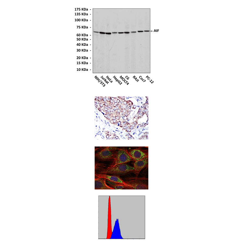
(Click to Enlarge) Top: Western blot detection of AIF proteins in various cell lysates using AIF Antibody. Middle, Upper: It also stains paraffin-embedded human breast cancer tissue in IHC analysis. Middle, Lower: This antibody stains NIH3T3 cells in confocal immunofluorescent testing (AIF Antibody: Green; Actin filaments: Red; DRAQ5 DNA Dye: Blue). Bottom: This antibody detects AIF proteins specifically in HepG2 cells by FACS assay (AIF Antibody: Blue; negative control: Red).
Details
Cat.No.: | CP10400 |
Antigen: | A raised against recombinant human AIF fragments expressed in E. coli. |
Isotype: | Mouse IgG1 |
Species & predicted species cross- reactivity ( ): | Human, Mouse, Rat |
Applications & Suggested starting dilutions:* | WB 1:1000 IP 1:50 IHC 1:50 - 1:200 ICC 1:50 - 1:200 FACS 1:50 - 1:200 |
Predicted Molecular Weight of protein: | 67 kDa |
Specificity/Sensitivity: | Detects AIF proteins in various cell lysate. |
Storage: | Store at -20°C, 4°C for frequent use. Avoid repeated freeze-thaw cycles. |
*Optimal working dilutions must be determined by end user.
Products
| Product | Size | CAT.# | Price | Quantity |
|---|---|---|---|---|
| Mouse AIF Antibody: Mouse AIF Antibody | Size: 100 ul | CAT.#: CP10400 | Price: $457.00 |
