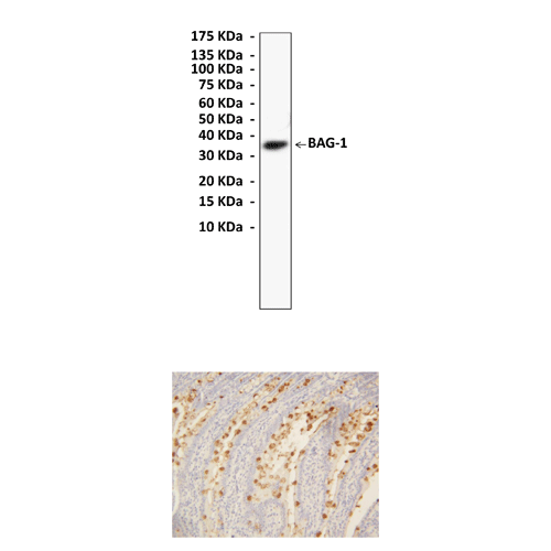
Description
BACKGROUND Bcl-2 associated athanogene (BAG-1) is a multifunctional protein which has a role in a range of cellular processes including apoptosis, cell survival, transcription, cell motility, and proliferation. It was first identified based on its ability to bind the anti-apoptotic protein Bcl-2 and to promote cell survival. Since its initial discovery as a Bcl-2-binding protein, however, BAG-1 has been reported to interact with and modulate the activities of other proteins.1 In human cells BAG-1 exists as three major isoforms (BAG-1S, BAG-1M and BAG-1L or p50, p46 and p36) derived by alternate translation initiation from a single mRNA. All BAG-1 isoforms contain a C-terminal, evolutionary conserved BAG domain and a central ubiquitin-like domain (ULD), but the larger isoforms have unique N-terminal extensions. The involvement of BAG-1 in the different cellular pathways can be in part explained by the sub-cellular compartmentalization of the three major BAG-1 isoforms. Thus the largest isoform p50 (BAG-1L), contains a nuclear localization sequence in the N-terminal extension and resides in the nucleus, whilst the most abundant form of the protein p36 (BAG-1S) is found predominately in the cytoplasm. BAG-1 interacts via its C-terminus (the BAG domain), with a large number of disparate proteins including. On the cell surface, it binds to the cytosolic domain of the growth factor receptors, enhancing the protection from cell death triggered by these receptors. In the cytosol, it binds to and modulates the function of BCL2, RAF-1, and subunits of the ubiquitinylation proteasome complex and Hsc70 and Hsp70. In the nucleus, it binds to a variety of nuclear hormone receptors and inhibits hormone-induced apoptosis.2
Given the role of BAG-1 in a number of different biological pathways, it is not surprising that deregulated BAG-1 expression is associated with carcinogenesis and moreover cells over-expressing BAG-1S are protected from apoptosis and are resistant to the effects of chemotoxic drugs. BAG-1 expression is known to protect cells from a wide range of apoptotic stimuli. Expression of BAG-1 is altered in various human malignancies, such as breast and lung cancer.3 BAG-1 expression is highly regulated and since the three major isoforms are generated from a single mRNA transcript translational regulation is thought to play a major role in the control of their expression.
Given the role of BAG-1 in a number of different biological pathways, it is not surprising that deregulated BAG-1 expression is associated with carcinogenesis and moreover cells over-expressing BAG-1S are protected from apoptosis and are resistant to the effects of chemotoxic drugs. BAG-1 expression is known to protect cells from a wide range of apoptotic stimuli. Expression of BAG-1 is altered in various human malignancies, such as breast and lung cancer.3 BAG-1 expression is highly regulated and since the three major isoforms are generated from a single mRNA transcript translational regulation is thought to play a major role in the control of their expression.
REFERENCES
1. Townsend, P.A et al: Biochim. Biophy. Acta 1603:83-98, 2003
2. Townsend, P.A et al: Int. J. Biochem. Cell Biol. 37;251-9, 2005
3. Cutress, R.I. et al: Br. J. Cancer 87:834-9, 2002
2. Townsend, P.A et al: Int. J. Biochem. Cell Biol. 37;251-9, 2005
3. Cutress, R.I. et al: Br. J. Cancer 87:834-9, 2002
Products are for research use only. They are not intended for human, animal, or diagnostic applications.
Details
Cat.No.: | CA1392 |
Antigen: | A short peptide from human BAG-1 carboxyl-terminal sequence. |
Isotype: | Rabbit IgG |
Species & predicted species cross- reactivity ( ): | Human, Rat |
Applications & Suggested starting dilutions:* | WB 1:500 - 1:1000 IP n/d IHC 1:50 - 1:100 ICC n/d FACS n/d |
Predicted Molecular Weight of protein: | 50, 45, 36 kDa |
Specificity/Sensitivity: | Detects endogenous BAG-1proteins without cross-reactivity with other family members. |
Storage: | Store at -20°C, 4°C for frequent use. Avoid repeated freeze-thaw cycles. |
*Optimal working dilutions must be determined by end user.
Products
| Product | Size | CAT.# | Price | Quantity |
|---|---|---|---|---|
| Rabbit BAG-1 Antibody: Rabbit BAG-1 Antibody | Size: 100 ul | CAT.#: CA1392 | Price: $302.00 |

