Product Sheet CA11007
Description
BACKGROUND Calbindin D belongs to the EF-hand family of calcium binding proteins and binds calcium ion with a fast association rate. It is widely expressed in Ca2+ transporting tissues such as epithelial-absorptive cells of the intestine and the distal tubular epithelial cells of the kidney. Calbindin-D was also found in the nervous system and it was suggested to co-localize with plasma membrane Ca2+ pumps. The function of Calbindin-D is dependent on the intracellular Ca2+ concentrations and it was shown that Ca2+ binding to Calbindin-D induced local structure changes around aromatic residues while no significant secondary structural changes were observed. In addition, Calbindin-D-containing neurons are thought to be related with memory, learning and long-term potentiation.1 It has been well known that Ca2+ plays a critical role in the light-mediated resetting of the circadian clock. Calbindin D influences Ca2+ buffering capacity of a cell, alters spatio-temporal aspects of intracellular Ca2+ signaling, and hence alters transmission of light information to the circadian clock in neurons of the suprachiasmatic nuclei (SCN).2 Moreover, Calbindin-D was also implicated in Parkinson’s disease (PD) since it was found that the dopaminergic neurons of the substantia nigra pars compacta (A9) expressing calbindin-D were more resistant to cell death than the neurons that do not express Calbindin-D in PD and in animal. In addition, alpha-Synuclein is a natively unfolded protein aggregation which is implicated in the pathogenesis of PD and several other neurodegenerative diseases, is known to interact with a great number of unrelated proteins. Some of these proteins, such as beta-synuclein and DJ-1, were shown to inhibit alpha-synuclein aggregation in vitro and in vivo therefore acting as chaperones. It was shown that Calbindin-D is also able to interact and co-aggregate with alpha-synuclein, significantly inhibiting alpha-synuclein fibrillation. Therefore, Calbindin-D28K can act as a chaperone, efficiently suppressing the process of alpha-synuclein fibril formation.3
REFERENCES
1. Lee, C.H. et al: Cell. Mol. Neurobiol. 29:665–72, 2009
2. Stadler, F. et al: Chronobiol. Int. 27:68-82, 2010
3. Zhou, W. et al: Cent. Eur. J. Biol. • 5: 11–20, 2010
2. Stadler, F. et al: Chronobiol. Int. 27:68-82, 2010
3. Zhou, W. et al: Cent. Eur. J. Biol. • 5: 11–20, 2010
Products are for research use only. They are not intended for human, animal, or diagnostic applications.
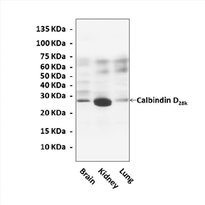
(Click to Enlarge) Western Blot detection of Calbindin D28k proteins in various rat tissue lysates using Calbindin D28k Antibody.
Details
Cat.No.: | CA11007 |
Antigen: | Short peptide from human Calbindin D28k sequence. |
Isotype: | Mouse IgG1 |
Species & predicted species cross- reactivity ( ): | Human, Mouse, Rat |
Applications & Suggested starting dilutions:* | WB 1:1000 IP n/d IHC 1:50 - 1:200 ICC n/d FACS n/d |
Predicted Molecular Weight of protein: | 28 kDa |
Specificity/Sensitivity: | Detects endogenous levels of Calbindin D28k proteins without cross-reactivity with other related proteins. |
Storage: | Store at -20°C, 4°C for frequent use. Avoid repeated freeze-thaw cycles. |
*Optimal working dilutions must be determined by end user.
Products
| Product | Size | CAT.# | Price | Quantity |
|---|---|---|---|---|
| Mouse Calbindin D28K Antibody: Mouse Calbindin D28K Antibody | Size: 100 ul | CAT.#: CA11007 | Price: $375.00 |