Product Sheet CA1201
Description
BACKGROUND Chemokines, a structurally related group of small chemotactic cytokines, are primary mediators of migration during the immune response. The principal immunological function of chemokines is two-fold: homeostatic (these are involved in basal leukocyte trafficking and/or the formation of the architecture of secondary lymphoid organs) and inflammatory (these are responsible for the recruitment of leukocytes into peripheral tissues in response to infection). CCL20, also known as macrophage inflammatory protein (MIP)-3alpha, Exodus-1, and LARC, induces migration through its cognate receptor, CCR6. However, CCL20-CCR6 does not constitute a monogamous binding pair, as recent studies have demonstrated that the beta-defensin group of antimicrobial peptides is able to bind and activate CCR6.1
CCR6 belongs to family A of G protein-coupled receptor superfamily. Cellular distribution of CCR6 is limited to leukocytes, with expression in mature lymphocytes, especially memory cells and immature dendritic cells (DCs) of particular lineages. In most cases it is absent from granulocytes (except activated neutrophils), monocytic cells, immature lymphocytes, and mature DC. Moreover, CCR6 is a selective marker for the identification of Th17 cells during an immune response. Furthermore, CCR6+ Th17 cells were present even in naïve mice and this population showed signs of activation in the steady state. During the acute phase of the immune response, the proportion of IL-17 producing Th cells in the lymph nodes decreased compared to the Th1 cell subpopulation.2 CCR6 has been reported to be of particular importance at mucosal sites. Compared with WT mice, Ccr6–/– mice have smaller Peyer patches, fewer associated M cells, and defective formation of isolated lymphoid follicles. Abnormalities in intestinal immune responses have been reported for Ccr6–/– mice in models of inflammatory bowel disease and during infection with rotavirus and Salmonella typhimurium. In the skin, Ccr6–/– mice have been reported to develop more or less severe contact hypersensitivity as well as diminished delayed-type hypersensitivity and graft-versus-host–mediated injury.3
CCR6 belongs to family A of G protein-coupled receptor superfamily. Cellular distribution of CCR6 is limited to leukocytes, with expression in mature lymphocytes, especially memory cells and immature dendritic cells (DCs) of particular lineages. In most cases it is absent from granulocytes (except activated neutrophils), monocytic cells, immature lymphocytes, and mature DC. Moreover, CCR6 is a selective marker for the identification of Th17 cells during an immune response. Furthermore, CCR6+ Th17 cells were present even in naïve mice and this population showed signs of activation in the steady state. During the acute phase of the immune response, the proportion of IL-17 producing Th cells in the lymph nodes decreased compared to the Th1 cell subpopulation.2 CCR6 has been reported to be of particular importance at mucosal sites. Compared with WT mice, Ccr6–/– mice have smaller Peyer patches, fewer associated M cells, and defective formation of isolated lymphoid follicles. Abnormalities in intestinal immune responses have been reported for Ccr6–/– mice in models of inflammatory bowel disease and during infection with rotavirus and Salmonella typhimurium. In the skin, Ccr6–/– mice have been reported to develop more or less severe contact hypersensitivity as well as diminished delayed-type hypersensitivity and graft-versus-host–mediated injury.3
REFERENCES
1. Yang, D. et al: Science 286: 525-8, 1999
2. Pötzl, J. et al: PLoS ONE 3:e2951, 2008
3. Hedrick, M.N. et al: J. Clin. Invest. 119:2317-29, 2009
2. Pötzl, J. et al: PLoS ONE 3:e2951, 2008
3. Hedrick, M.N. et al: J. Clin. Invest. 119:2317-29, 2009
Products are for research use only. They are not intended for human, animal, or diagnostic applications.
Details
Cat.No.: | CA1201 |
Antigen: | Short peptide from human CCR6 sequence. |
Isotype: | Rabbit IgG |
Species & predicted species cross- reactivity ( ): | Human, Rat |
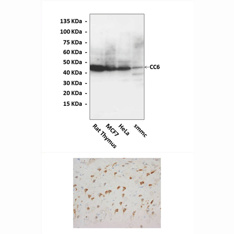
Applications & Suggested starting dilutions:* | WB 1:1000 IP n/d IHC 1:50 - 1:200 ICC n/d FACS n/d |
Predicted Molecular Weight of protein: | 46 kDa |
Specificity/Sensitivity: | Detects endogenous levels of CCR6 proteins without cross-reactivity with other related proteins. |
Storage: | Store at -20°C, 4°C for frequent use. Avoid repeated freeze-thaw cycles. |
*Optimal working dilutions must be determined by end user.
Products
| Product | Size | CAT.# | Price | Quantity |
|---|---|---|---|---|
| Rabbit CCR6 Antibody: Rabbit CCR6 Antibody | Size: 100 ul | CAT.#: CA1201 | Price: $375.00 |

