Product Sheet CP10065
Description
BACKGROUND Cytokeratins (CKs) are the largest class of cytoskeletal intermediate filament proteins. There are 20 different Cytokeratin proteins (assigned numbers 1-20), which are structural markers specific for epithelial cells, and are expressed specifically in different types of epithelial tissue. Cytokeratins are primarily insoluble molecules that play an important role in cellular mechanics, including: cell shape, motility, division and cell-cell contact.
There are two types of Cytokeratins. Type I (9–20) Cytokeratins are relatively acidic and are of small molecular weight (40–56.5 kDa). Type II (1–8) Cytokeratins are relatively basic-neutral and of larger molecular weight (53–67 kDa). Expression of CK in epithelia is dependent on types (simple or stratified), and differentiation pattern of epithelia. CK can be subdivided into markers of cornification (CK 1, 2, 10, and 11), stratification (CK4, 13), basal cells (CK 5, 14, 15, and 17), simple cells (CK 7, 8, 18, and 19), and hyperproliferation (CK6, 16). Although CKs are expressed in specific combinations in different epithelia, the number of permutations is limited, and thus insufficient to endow every epithelium with its own unique CK profile.1
Cytokeratin 18 is expressed at high levels by many cells found in single layer epithelial tissues. This protein, together with CK8, exhibits resistance features in response to stress and to apoptosis. It is often used together with CK8 and CK19 to differentiate cells of epithelial origin from hematopoietic cells in tests that enumerate circulating tumor cells in blood.2 A neo-epitope in CK18 cleaved at Asp396, termed M30-antigen, becomes available at an early caspase cleavage event during apoptosis, and is not detectable in vital or necrotic cells. By contrast, the cytosolic pool of uncleaved CK18 (also termed M65-antigen) is released from cells during necrosis. These findings suggest that different forms of CK18 in patient sera (M30-antigen for apoptosis and M65-antigen for necrosis) could be used to examine different cell death modes in vivo.3 Mutations in CK18 gene have been linked to cryptogenic cirrhosis.
REFERENCES
1. Makino T et al.: Br. J. Cancer 101:1298-1306 (2009)
2. Allard WJ et al.: Clin. Cancer Res. 10:6897-6904 (2004).
3. Vos MB et al.: J. Pediatric Gastroenterol. Nutri. 47:481-485 (2008).
2. Allard WJ et al.: Clin. Cancer Res. 10:6897-6904 (2004).
3. Vos MB et al.: J. Pediatric Gastroenterol. Nutri. 47:481-485 (2008).
Products are for research use only. They are not intended for human, animal, or diagnostic applications.
(Click to Enlarge)
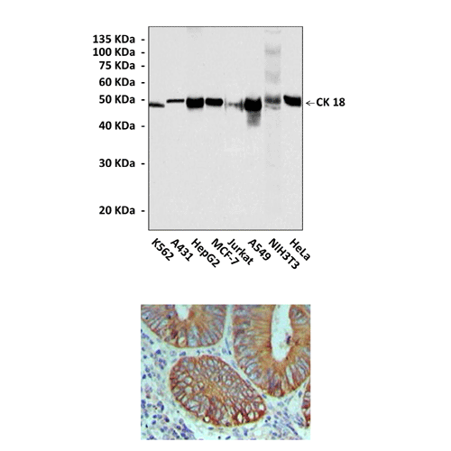
Top: Western Blot detection of endogenous Cytokeratin proteins from various cell lysates using Cytokeratin 18 Mouse Monoclonal Antibody (CK18). Bottom: This antibody stains paraffin-embedded human colon cancer tissue in immunohistochemical analysis.
Details
Cat.No.: | CP10065 |
Antigen: | Recombinant human Cytokeratin 18 fragment containing its carboxyl terminal sequence. |
Isotype: | Mouse IgG2b |
Species & predicted species cross- reactivity ( ): | Human, Mouse, Rat |
Applications & Suggested starting dilutions:* | WB 1:500 - 1:2000 IP n/d IHC (Paraffin) 1:200 - 1:1000 ICC n/d FACS n/d |
Predicted Molecular Weight of protein: | 48 kDa |
Specificity/Sensitivity: | Detects endogenous levels of Cytokeratin18 protein in normal cell lysates without cross-reactivity with other family members |
Storage: | Store at -20°C, 4°C for frequent use. Avoid repeated freeze-thaw cycles. |
*Optimal working dilutions must be determined by end user.
Products
| Product | Size | CAT.# | Price | Quantity |
|---|---|---|---|---|
| Mouse Cytokeratin 18 Antibody: Mouse Cytokeratin 18 Antibody | Size: 100 ul | CAT.#: CP10065 | Price: $472.00 |