Product Sheet CP10287
Description
BACKGROUND The Ets family of transcription factors, of which there are now almost 30 members, are characterized by a winged helix-turn-helix DNA-binding domain termed the ‘Ets’ domain. ETS family members bind to palindromic ETS-binding sites (EBS) composed of a 5’-GGA(A/T)-3’ consensus core sequence and regulate expression of genes involved in proliferation (Myc), invasion (matrix metalloproteinase (MMP)-1, MMP-3, MMP-9 and urokinase plasminogen activator), and angiogenesis (VEGF receptor 1).1 Ets factors can be subcategorized into subfamilies based on DNA and protein sequence homology. For example, Elf-1, Nerf-2, and Mef are highly related and belong to one subfamily. Similarly, Fli1 and ERG constitute another subfamily, as do Ets1 and Ets2. The subfamily members often have overlapping functions. From an evolutionary standpoint, this built-in redundancy may be protective against the effects of genetic mutations of the individual family members for critical developmental processes such as vasculogenesis.2
Ets1, the founding member of the Ets transcription factor family, is a cellular homologue of the avian erythroblastosis E26 viral oncogene that contains a conserved 85 amino acid ETS DNA binding domain. The DNA binding activity of Ets1 is controlled by kinases and transcription factors. Some transcription factors, such as AML-1, regulate Ets1 by targeting its autoinhibitory module. Others, such as Pax-5, alter Ets1 DNA binding properties. Ets1 harbors two phosphorylation sites, threonine-38 and an array of serines within the exon VII domain. Phosphorylation of threonine-38 by ERK1/2 activates Ets1, whereas phosphorylation of the exon VII domain by CaMKII or MLCK inhibits Ets1 DNA binding activity. Ets1 is expressed by numerous cell types. Ets1 is thought to regulate the expression of proteases such as urokinase plasminogen activator (uPA) and various members of the matrix metalloprotease family (MMP1, MMP2, MMP3, MMP9 and MMP13). Expression of these proteases is likely required for tumor cells to degrade the surrounding extracellular matrix, invade nearby tissues and eventually metastasize to distant sites. In addition, to these proteases, Ets1 is also implicated in upregulation of a wide-variety of genes that may promote tumorigenesis including genes that control cellular proliferation or survival (bax, bcl-2, caspase-1, c-myc, CDK11, Fas ligand, GADD153, JunB, mdm2, p16, p21 and p53) and genes involved in responses to a hypoxic environment. Furthermore, Ets1 has been reported to directly interact with the tumor suppressor protein p53 and modulate its transcriptional activity. In haemotopoietic cells, Ets1 contributes to the regulation of cellular differentiation. In a variety of other cells, including endothelial cells, vascular smooth muscle cells and epithelial cancer cells, Ets1 promotes invasive behavior. In tumors, Ets1 expression is indicative of poorer prognosis.3 From a therapeutic standpoint, recent studies support an important role for Ets factors as critical transcriptional regulators of tumor angiogenesis. Local administration of a dominant-negative form of Ets1, encoding the DNA binding domain, significantly blocked tumor angiogenesis and tumor growth. With recent advances in drug discovery, the identification and/or development of small molecule inhibitors of subfamilies of ETS factors may now be possible by taking advantage of structural similarities among the subfamilies in the DNA binding domain or other functionally conserved regions of the proteins.4
Ets1, the founding member of the Ets transcription factor family, is a cellular homologue of the avian erythroblastosis E26 viral oncogene that contains a conserved 85 amino acid ETS DNA binding domain. The DNA binding activity of Ets1 is controlled by kinases and transcription factors. Some transcription factors, such as AML-1, regulate Ets1 by targeting its autoinhibitory module. Others, such as Pax-5, alter Ets1 DNA binding properties. Ets1 harbors two phosphorylation sites, threonine-38 and an array of serines within the exon VII domain. Phosphorylation of threonine-38 by ERK1/2 activates Ets1, whereas phosphorylation of the exon VII domain by CaMKII or MLCK inhibits Ets1 DNA binding activity. Ets1 is expressed by numerous cell types. Ets1 is thought to regulate the expression of proteases such as urokinase plasminogen activator (uPA) and various members of the matrix metalloprotease family (MMP1, MMP2, MMP3, MMP9 and MMP13). Expression of these proteases is likely required for tumor cells to degrade the surrounding extracellular matrix, invade nearby tissues and eventually metastasize to distant sites. In addition, to these proteases, Ets1 is also implicated in upregulation of a wide-variety of genes that may promote tumorigenesis including genes that control cellular proliferation or survival (bax, bcl-2, caspase-1, c-myc, CDK11, Fas ligand, GADD153, JunB, mdm2, p16, p21 and p53) and genes involved in responses to a hypoxic environment. Furthermore, Ets1 has been reported to directly interact with the tumor suppressor protein p53 and modulate its transcriptional activity. In haemotopoietic cells, Ets1 contributes to the regulation of cellular differentiation. In a variety of other cells, including endothelial cells, vascular smooth muscle cells and epithelial cancer cells, Ets1 promotes invasive behavior. In tumors, Ets1 expression is indicative of poorer prognosis.3 From a therapeutic standpoint, recent studies support an important role for Ets factors as critical transcriptional regulators of tumor angiogenesis. Local administration of a dominant-negative form of Ets1, encoding the DNA binding domain, significantly blocked tumor angiogenesis and tumor growth. With recent advances in drug discovery, the identification and/or development of small molecule inhibitors of subfamilies of ETS factors may now be possible by taking advantage of structural similarities among the subfamilies in the DNA binding domain or other functionally conserved regions of the proteins.4
REFERENCES
1. Lelièvre, E. et al: Int. J. Biochem. Cell. Biol. 33:391-407, 2001
2. Oettgen, P.: Blood 114:934-5, 2009
3. Dittmer, J.: Mol. Cancer 2:29, 2003
4. Davidson, B. et al: Cancer Metastasis Rev. 22:103-15, 2003
2. Oettgen, P.: Blood 114:934-5, 2009
3. Dittmer, J.: Mol. Cancer 2:29, 2003
4. Davidson, B. et al: Cancer Metastasis Rev. 22:103-15, 2003
Products are for research use only. They are not intended for human, animal, or diagnostic applications.

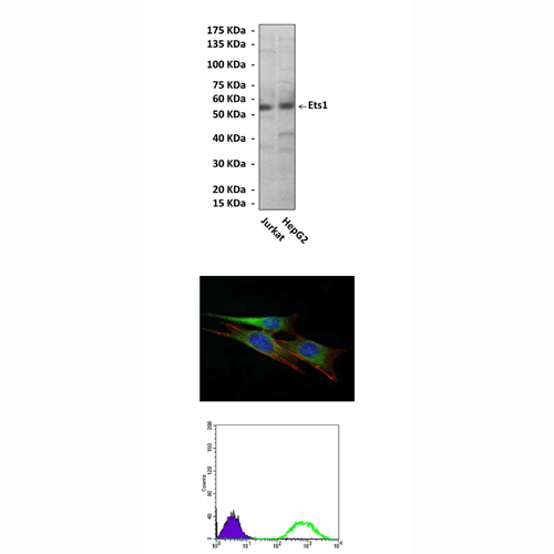
(Click to Enlarge) Top: Western Blot detection of Ets1 proteins in Jurkat and HepG2 cell lysates using Ets1 Antibody. Middle: This antibody stains NIH3T3 cells in confocal immunofluorescent analysis (Ets1 Antibody: Green; Actin filaments: Red; DRAQ5 DNA Dye: Blue). Bottom: It also specifically reacts with Ets1 proteins in Jurkat cells by FACS testing (Ets1 Antibody: Green; control; Purple).
Details
Cat.No.: | CP10287 |
Antigen: | Raised against recombinant human Ets1 fragments expressed in E. coli. |
Isotype: | Mouse IgG1 |
Species & predicted species cross- reactivity ( ): | Human, Mouse, Rat |
Applications & Suggested starting dilutions:* | WB 1:1000 IP n/d IHC n/d ICC 1:50 - 1:200 FACS 1:50 - 1:200 |
Predicted Molecular Weight of protein: | 55 kDa |
Specificity/Sensitivity: | Detects endogenous Ets1 proteins without cross-reactivity with other family members. |
Storage: | Store at -20°C, 4°C for frequent use. Avoid repeated freeze-thaw cycles. |
*Optimal working dilutions must be determined by end user.
Products
| Product | Size | CAT.# | Price | Quantity |
|---|---|---|---|---|
| Mouse Ets1 Antibody: Mouse Ets1 Antibody | Size: 100 ul | CAT.#: CP10287 | Price: $413.00 |
