Product Sheet CP10357
Description
BACKGROUND The hexokinases (HKs) utilize Mg-ATP as a phosphoryl donor to catalyze the first step of intracellular glucose metabolism, the conversion of glucose to glucose- 6-phosphate. ). Thus, Hexokinase initiates all major pathways of intracellular glucose utilization Four hexokinase isoenzymes have been identified, including hexokinase I (HK I), hexokinase II (HK II), hexokinase III (HK III) and hexokinase IV (HK IV, also designated glucokinase or GCK). Accumulating evidence indicates that the mitochondrially bound isoforms of hexokinase, HK-I and HK-II, play pivotal roles in promoting cell growth and survival in rapidly growing, highly glycolytic tumors. They couples glycolysis to oxidative phosphorylation by interacting with mitochondria, thus acting as a metabolic sensor. In highly glycolytic, i.e. extremely aggressive tumours, mitochondrial HK II activity is increased and fosters cell growth in the hypoxic conditions of neoplastic mass accrual by enhancing glycolysis, which becomes independent of oxygen availability (the Warburg effect). With the re-emergence and acceptance of both the “Warburg effect” as a prominent phenotype of most clinical cancers, and “metabolic targeting” as a rational therapeutic strategy, a number of laboratories are focusing on metabolite entry or exit steps. One remarkable success story is the use of the small molecule 3-bromopyruvate (3-BP) that selectively enters and destroys the cells of large tumors in animals by targeting both HK-2 and the mitochondrial ATP synthasome. This leads to very rapid ATP depletion and tumor destruction without harm to the animals.1 Furthermore, mitochondrial HK II plays an important role in maintaining the integrity of the outer mitochondrial membrane (OMM), thus preventing the release of key apoptogenic molecules from the intermembrane space. Reports indicate that nutrients, via the survival kinase Akt, promote HK II binding to the voltage-dependent anion channel [VDAC], a protein that allows for the movement of small metabolites across the OMM. Release of HK II transmits a potent death signal that is elicited when GSK3β, a kinase inhibited by Akt, phosphorylates a putative HK docking site on VDAC. It has been documented that inhibition of GSK-3β results in GSK-3β-mediated resistance to oxidant stress. When associated to HK II, VDAC interacts with antiapoptotic Bcl-2 family members. These might be competed away from VDAC by the pro-apoptotic Bax/Bak proteins after deprivation of growth factors, leading to the formation of a conduit on the OMM fit for the release of apoptogenic proteins. Alternatively, the HK/VDAC interaction could transmit molecular changes to proteins of the inner mitochondrial membrane (IMM), resulting in permeability transition pore (PTP) regulation. The PTP is an IMM channel whose opening elicits depolarization, matrix swelling, and consequently cristae unfolding and breaches in the OMM that are pervious to proteins. A distinction between these two possible scenarios would have important consequences both on the comprehension of apoptosis dysregulation in tumors and on the design of therapeutic approaches targeted to selectively eliminate neoplastic cells. It was shown that cell death mediated by HK detachment from mitochondria is selectively sensitive to modulators of the PTP. Moreover, VDAC is dispensable for carrying out this process.2 In addition, It was shown that mTORC1 hyperactivity inhibits serum deprivation-induced apoptosis via increased hexokinase II and GLUT1 Expression, sustained Mcl-1 expression, and glycogen synthase kinase 3β inhibition.3 Recently, it was shown that hexokinase II shuttled between cytoplasma and nuclei in tumor cells. Regulated translocation of HXKII to the nucleus of mammalian cells could represent a previously unknown glucose-sensing mechanism.4
REFERENCES
1. Mathupala, S.P. et al: Sem. Cancer Biol. 19:17-24, 2009
2. Chiara, F. et al: PloS ONE 3:e1852, 2008
3. Bhaskar,P.T. et al: Mol. Cell. Biol. 29:5136-47, 2009
4. Neary, C.L. & Pastorino, J.G.: Biochem. Biophy. Res. Commun. 394:1705-81, 2010
2. Chiara, F. et al: PloS ONE 3:e1852, 2008
3. Bhaskar,P.T. et al: Mol. Cell. Biol. 29:5136-47, 2009
4. Neary, C.L. & Pastorino, J.G.: Biochem. Biophy. Res. Commun. 394:1705-81, 2010
Products are for research use only. They are not intended for human, animal, or diagnostic applications.

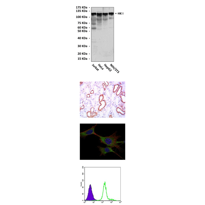
(Click to Enlarge) Top: Western blot detection of hexokinase I proteins in various cell lysates using Hexokinase I Antibody. Middle upper: This antibody stains paraffin-embedded human kidney tissue in immunohistochemical analysis. Middle lower: It also stains NIH3T3 cells in confocal immunofluorescent testing (Hexokinase I Antibody: Green; Actin filaments: Red; DRAQ5 DNA Dye: Blue). Bottom: It also specifically react with Hexokinase II proteins in K562 cells by FACS assay (Hexokinase II Antibody: Green; control: purple).
Details
Cat.No.: | CP10357 |
Antigen: | Raised against recombinant human HK I fragments expressed in E. coli. |
Isotype: | Mouse IgG1 |
Species & predicted species cross- reactivity ( ): | Human, Mouse |
Applications & Suggested starting dilutions:* | WB 1:1000 IP n/d IHC 1:50 - 1:200 ICC 1:50 - 1:200 FACS 1:50 - 1:200 |
Predicted Molecular Weight of protein: | 120 kDa |
Specificity/Sensitivity: | Detects HK I proteins in various cell lysate. |
Storage: | Store at -20°C, 4°C for frequent use. Avoid repeated freeze-thaw cycles. |
*Optimal working dilutions must be determined by end user.
Products
| Product | Size | CAT.# | Price | Quantity |
|---|---|---|---|---|
| Mouse Hexokinase I Antibody: Mouse Hexokinase I Antibody | Size: 100 ul | CAT.#: CP10357 | Price: $413.00 |
