Product Sheet CP10438
Description
BACKGROUND Members of the superfamily of voltage-gated cation channels open and close in response to changes in the voltage across the cell membrane to generate electrical currents that underlie neuronal signaling and skeletal and cardiac muscle contraction. The voltage-gated openings of these channels are controlled by intrinsic voltage sensors, including several positively charged residues in the fourth transmembrane (S4) segment of the channel, which move in response to changes in the membrane voltage. The voltage sensor movements trigger opening of the channel gate through a coupling mechanism that is not well-understood, and which may vary between specific channels and channel complexes. A fundamental understanding of how voltage sensors move and how this movement couples to channel opening is important to understand how these channels function during physiological and pathophysiological conditions and how mutation of these channels and their associated accessory subunits causes disease. One channel whose gating properties are of particular interest is the IKs potassium channel, a slowly activating channel that is essential to normal cardiac function. That the unique biophysical properties of the IKs channel are important for normal cardiac physiology is evidenced by multiple pathophysiological clinical phenotypes associated with mutations that change the biophysical and regulatory properties of the IKs channel, such as long QT syndrome, short QT syndrome, and familial atrial fibrillation.1 The IKs channel consists of an α-subunit (KCNQ1, also known as Kv7.1) and an accessory β-subunit (KCNE1). KCNQ1 belongs to the canonical voltage-gated potassium channel family, forming a homotetrameric channel with a central pore domain and four peripheral voltage-sensing domains. KCNE1 is a small (129-amino acid) single-pass transmembrane protein. Although four KCNQ1 subunits assemble to form functional tetrameric voltage-gated channels, the biophysical and regulatory properties of the KCNQ1 channel when expressed alone are completely distinct from IKs currents: KCNQ1 homomeric channels activate and deactivate rapidly and begin to open at voltages more negative than those that activate IKs channels. Coassembly of KCNE1 with KCNQ1 drastically slows the activation kinetics, shifts the voltage dependence of activation, slows deactivation, and increases single-channel conductance, thereby reproducing the critical properties of the native IKs channel.2
The KCNQ1 gene has a total of 17 exons, spans 404 kb of chromosome sequence and is located on chromosome 11p15.5. KCNQ1 codes for the pore-forming alpha subunit of the voltage-gated K+ channel (KvLQT1) that is highly expressed in the heart. This channel plays an important role in controlling repolarization of the ventricles, where its primary function is to limit action potential prolongation during sympathetic stimulation. Mutations in KCNQ1 have been described to lead to cardiac long QT syndrome, Jervell and Lange-Nielsen syndrome, which are associated with cardiac conduction abnormalities and hearing loss. KCNQ1 is also expressed to lesser extent in the pancreas, placenta, lung, liver, kidney, brain, and adipose tissue. In addition, KCNQ1 is expressed in vitro in insulin-secreting cell lines. Insulin secretion from pancreatic β cells is regulated by complex interplay between KATP channels and Kv- channels and voltage-dependent Ca++ channels. Ionic mechanisms at KATP and Kv- channels are primarily important in triggering and maintaining glucose-stimulated insulin secretion and inhibition of this potassium channel has been shown to significantly increase insulin secretion. It was shown that that the variation within the KCNQ1 locus confers a significant risk to type II diabetes (T2D).3
The KCNQ1 gene has a total of 17 exons, spans 404 kb of chromosome sequence and is located on chromosome 11p15.5. KCNQ1 codes for the pore-forming alpha subunit of the voltage-gated K+ channel (KvLQT1) that is highly expressed in the heart. This channel plays an important role in controlling repolarization of the ventricles, where its primary function is to limit action potential prolongation during sympathetic stimulation. Mutations in KCNQ1 have been described to lead to cardiac long QT syndrome, Jervell and Lange-Nielsen syndrome, which are associated with cardiac conduction abnormalities and hearing loss. KCNQ1 is also expressed to lesser extent in the pancreas, placenta, lung, liver, kidney, brain, and adipose tissue. In addition, KCNQ1 is expressed in vitro in insulin-secreting cell lines. Insulin secretion from pancreatic β cells is regulated by complex interplay between KATP channels and Kv- channels and voltage-dependent Ca++ channels. Ionic mechanisms at KATP and Kv- channels are primarily important in triggering and maintaining glucose-stimulated insulin secretion and inhibition of this potassium channel has been shown to significantly increase insulin secretion. It was shown that that the variation within the KCNQ1 locus confers a significant risk to type II diabetes (T2D).3
REFERENCES
1. Lundby, A. et al: Heart Rhythm 7:708-13, 2010
2. Osteen, J.D. et al: Proc. Natl. Acad. Sci. USA 107:22710–5, 2010
3. Been, J.F. et al: BMC Med. Genet. 12:18, 2011
2. Osteen, J.D. et al: Proc. Natl. Acad. Sci. USA 107:22710–5, 2010
3. Been, J.F. et al: BMC Med. Genet. 12:18, 2011
Products are for research use only. They are not intended for human, animal, or diagnostic applications.
Details
Cat.No.: | CP10438 |
Antigen: | Recombinant human KCNQ1 fragments expressed in E. coli. |
Isotype: | Mouse IgG1 |
Species & predicted species cross- reactivity ( ): | Human, Mouse, Rat |
Applications & Suggested starting dilutions:* | WB 1:1000 IP n/d IHC 1:50 - 1:200 ICC 1:50 - 1:200 FACS 1:50 - 1:200 |
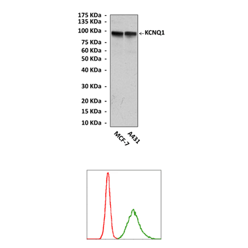
Predicted Molecular Weight of protein: | 95 kDa |
Specificity/Sensitivity: | Detects endogenous KCNQ1 proteins without cross-reactivity with other family members. |
Storage: | Store at -20°C, 4°C for frequent use. Avoid repeated freeze-thaw cycles. |
*Optimal working dilutions must be determined by end user.
Products
| Product | Size | CAT.# | Price | Quantity |
|---|---|---|---|---|
| Mouse KCNQ1 Antibody: Mouse KCNQ1 Antibody | Size: 100 ul | CAT.#: CP10438 | Price: $457.00 |

