Product Sheet CP10335
Description
BACKGROUND Keratins belong to the family of intermediate filament proteins that are specifically expressed in epithelia. They have a remarkable ability to polymerize into 10nm filaments without the participation of auxiliary proteins. They comprise a total of about 30 genes (including those of hair and nails, the trichocytic keratins) grouped into two types; type I are smaller (40–56.5kDa) and acidic (pI<7.0), whereas type II are larger (53–67kDa) and basic/neutral (pI ≥7.0). The type I keratins include K9–K20 and the type II include K1–K8. The amino acid sequence of keratins is highly conserved in the central rod domain of the polypeptides and forms an alpha-helical structure. During filament assembly, two keratin polypeptides, one of each type, first form a parallel heterodimer, in which the rod domains assemble into coiled-coil, which then undergoes further associations with other dimers to produce tetramers. The association of tetramers produces protofilaments and finally mature filaments. Keratins are often expressed in specific pairs in an epithelial tissue that are unique to that differentiation pathway. The keratinocytes in the mitotically active basal layer always express K5 and K14, and upon commitment to differentiation the basal keratinocytes exit from the cell-cycle and move into the suprabasal compartment. The migrating keratinocytes downregulate K5/K14 transcription and activate expression of a new set of keratin pairs that vary among stratified tissues. In cornified epithelia, such as those covering skin and gingivae, the differentiating keratinocytes express K1/K10, whereas in noncornified squamous epithelia expression of K4/K13, is induced and in cornea the migrating keratinocytes activate K3/K12 expression. The K6/K16 pair is constitutively expressed in the outer root sheath of hair follicles and in mucosal epithelia of oral cavity, esophagus, and female genital tract; however, in epidermal hyperproliferation, such as in psoriasis and during wound healing, expression of this pair is activated.1
K15 is a type I keratin that does not appear to have a natural type II expression partner. Earlier studies using radioactive in situ hybridization have shown that the K15 mRNA is expressed in all layers of stratified epithelia. The conclusion drawn from these studies was that the K15 expression starts in the basal layer but is independent of the vertical differentiation of migrating keratinocytes. Recent studies, however, have shown that K15 is not expressed in the suprabasal layer but instead is specifically localized in the basal keratinocytes.2 It is suggested that keratin 15 is a good marker for the stem cells in the human hair follicle bulge areas. Despite the fact that K15 is regarded as specific for basal keratinocytes, its distribution in normal and epithelial-related diseases has not been investigated in detail. Expression of K15 also suppressed by tumor necrosis factor alpha (TNF-alpha) and to a lesser extent by epidermal growth factor (EGF) and keratinocyte growth factor (KGF).3 Thus, K15 is excluded from the activated keratinocytes of the hyperthickened wound epidermis, possibly as a result of increased growth factor expression in injured skin. In addition, K15 and K17 proteolysis was observed during staurosporine-induced apoptosis and anoikis (anchorage-dependent apoptosis) as well and was shown to be caspase-dependent.4
REFERENCES
1. Waseem, A. et al: J. Investigat. Dermatol. 112:362–369, 1999
2. Ohyama, M. et al: J. Clin. Invest. 116:249-60, 2006
3. Werner, S. et al: Exp. Cell Res. 254:80-90, 2000
4. Badock, V. et al: Cell Death Diff. 8:308-315, 2001
2. Ohyama, M. et al: J. Clin. Invest. 116:249-60, 2006
3. Werner, S. et al: Exp. Cell Res. 254:80-90, 2000
4. Badock, V. et al: Cell Death Diff. 8:308-315, 2001
Products are for research use only. They are not intended for human, animal, or diagnostic applications.

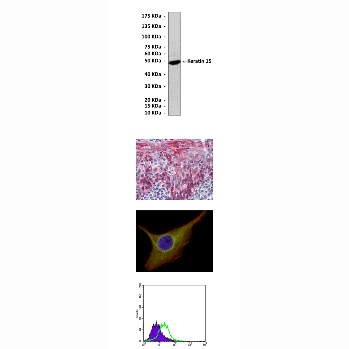
(Click to Enlarge) Top: Western Blot detection of Keratin 15 proteins in A431 cell lysate using Keratin 15 Antibody. Middle, upper: This antibody stains paraffin-embedded human tonsil tissue in immunohistochemical analysis. Middle, lower: It also stains PACN-1 cells in confocal immunofluorescent analysis (Keratin antibody: Green; Actin filaments: Red; DRAQ% DNA dye: Blue). Bottom: It also specifically reacts with Keratin 15 protein in PACN-1 cells in FACS testing (Keratin 15 antibody: Green; normal mouse IgG control: Blue).
Details
Cat.No.: | CP10149 |
Antigen: | Purified human Keratin 15 fragments expressed in E. coli. |
Isotype: | Mouse IgG2 |
Species & predicted species cross- reactivity ( ): | Human |
Applications & Suggested starting dilutions:* | WB 1:1000 IP n/d IHC 1:200 ICC 1:200 FACS 1:200 |
Predicted Molecular Weight of protein: | 49 kDa |
Specificity/Sensitivity: | Detects endogenous Keratin 15 proteins without cross-reactivity with other family members. |
Storage: | Store at -20°C, 4°C for frequent use. Avoid repeated freeze-thaw cycles. |
*Optimal working dilutions must be determined by end user.
Products
| Product | Size | CAT.# | Price | Quantity |
|---|---|---|---|---|
| Mouse Keratin 15 Antibody: Mouse Keratin 15 Antibody | Size: 100 ul | CAT.#: CP10335 | Price: $413.00 |
