Product Sheet CP10405
Description
BACKGROUND Cytokeratins (CKs) are the largest class of cytoskeletal intermediate filament proteins. There are 20 different Cytokeratin proteins (assigned numbers 1-20), which are structural markers specific for epithelial cells, and are expressed specifically in different types of epithelial tissue. Cytokeratins are primarily insoluble molecules that play an important role in cellular mechanics, including: cell shape, motility, division and cell-cell contact.
There are two types of Cytokeratins. Type I (9–20) Cytokeratins are relatively acidic and are of small molecular weight (40–56.5 kDa). Type II (1–8) Cytokeratins are relatively basic-neutral and of larger molecular weight (53–67 kDa). Expression of CK in epithelia is dependent on types (simple or stratified), and differentiation pattern of epithelia. CK can be subdivided into markers of cornification (CK 1, 2, 10, and 11), stratification (CK4, 13), basal cells (CK 5, 14, 15, and 17), simple cells (CK 7, 8, 18, and 19), and hyperproliferation (CK6, 16). Although CKs are expressed in specific combinations in different epithelia, the number of permutations is limited, and thus insufficient to endow every epithelium with its own unique CK profile.1
Cytokeratin 19 is a type I cytokeratin. Unlike its related family members, this smallest known acidic cytokeratin is not paired with a basic cytokeratin in epithelial cells. It is specifically found in the periderm, the transiently superficial layer that envelops the developing epidermis. CK19 is often used together with CK8 and CK18 to differentiate cells of epithelial origin from hematopoietic cells in tests that enumerate circulating tumor cells in blood.2 CK19 is stably and abundantly expressed in epithelial tumors, but not in mesenchymal hemopoietic cells, and has been successfully used as a marker for the detection of tumor cells in the bone marrow, lymph nodes, and peripheral blood by immunohistochemistry and RT-PCR.3
REFERENCES
1. Makino T et al.: Br. J. Cancer 101:1298-1306 (2009).
2. Allard WJ et al.: Clin. Cancer Res. 10:6897-6904 (2004).
3. Stathopoulou A et al.: Clin. Cancer Res. 9:5145-5151, (2003).
2. Allard WJ et al.: Clin. Cancer Res. 10:6897-6904 (2004).
3. Stathopoulou A et al.: Clin. Cancer Res. 9:5145-5151, (2003).
Products are for research use only. They are not intended for human, animal, or diagnostic applications.
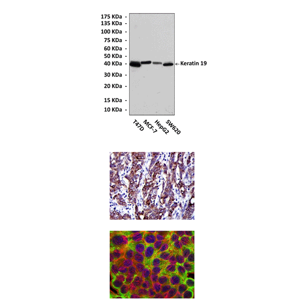
(Click to Enlarge) Top: Western blot detection of keratin 19 proteins in various cell lysates using keratin 19 Antibody. Middle: This antibody stains HepG2 cells in confocal immunofluorescent testing (keratin 19 antibody: Green; Actin filaments: Red; DRAQ5 DNA Dye: Blue). Bottom: It also stains paraffin-embedded human cervical cancer tissue in IHC analysis.
Details
Cat.No.: | CP10405 |
Antigen: | Raised against recombinant human keratin 19 fragments expressed in E. coli. |
Isotype: | Mouse IgG1 |
Species & predicted species cross- reactivity ( ): | Human, Mouse, Rat |
Applications & Suggested starting dilutions:* | WB 1:1000 IP n/d IHC 1:50 - 1:200 ICC 1:50 - 1:200 FACS n/d |
Predicted Molecular Weight of protein: | 40 kDa |
Specificity/Sensitivity: | Detects keratin 19 proteins in various cell lysate. |
Storage: | Store at -20°C, 4°C for frequent use. Avoid repeated freeze-thaw cycles. |
*Optimal working dilutions must be determined by end user.
Products
| Product | Size | CAT.# | Price | Quantity |
|---|---|---|---|---|
| Mouse Keratin 19 Antibody: Mouse Keratin 19 Antibody | Size: 100 ul | CAT.#: CP10405 | Price: $457.00 |