Product Sheet CC10007
Description
BACKGROUND Phosphohexose isomerase (PHI; D-glucose-6-phosphate ketol-isomerase; EC 5.3.1.9) is also known as glucosephosphate isomerase (GPI) and phosphoglucose isomerase (PGI). It is a housekeeping cytosolic enzyme of sugar metabolism that plays a key role in both glycolysis and gluconeogenesis pathways, catalyzing the interconversion of glucose 6-phosphate and fructose 6-phosphate, the second step of the Embden-Meyerhof glycolytic pathway. And This enzyme is universally distributed among Eukaryotes, bacteria, and some Archaea. There is evidence that phosphoglucose isomerase behaves extracellularly as a cytokine. It is produced and secreted by white blood cells, and acts to regulate the growth of several different cell types.1 Molecular cloning and sequencing have identified PGI as an autocrine motility factor (AMF) found to be a major cell motility–stimulating factor associated with cancer development and progression.2 Of note, aberrations in PGI expressions or activities due to mutations or deletions in PGI are of significant clinical importance because mutations in PGI lead to hereditary nonspherocytic hemolytic anemia disease.3 In clinical cancer pathology, the presence of PGI/AMF in the serum and urine is of prognostic value indicating cancer progression. The levels of PGI/AMF and its cell surface receptor gp78/AMFR expressions are associated with the pathologic stage, grade, and degree of tumor penetration to surrounding tissues marking a poor prognosis.4
REFERENCES
1. Kim JW & Dang CV: Trends Biochem. Sci. 30:142–502, 2005.
2. Niinaka Y et al.: Cancer Res 58:2667–74, 1998.
3. Kanno H et al.: Blood 88:2321-2325, 1996.
4. Gomm SA et al.: Br. J. Cancer 58:797–804, 1988.
2. Niinaka Y et al.: Cancer Res 58:2667–74, 1998.
3. Kanno H et al.: Blood 88:2321-2325, 1996.
4. Gomm SA et al.: Br. J. Cancer 58:797–804, 1988.
Products are for research use only. They are not intended for human, animal, or diagnostic applications.

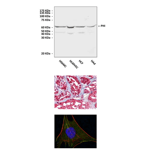
(Click to Enlarge) Top: Various primary cell lysates were subjected to Western Blot analysis using Phosphohexose Isomerase antibody. Middle: Immunohistochemical analysis of paraffin-embedded human Kidney tissues using PHI Antibody. Bottom: Immunofluorescence analysis of L-02 cells using PHI Antibody (PHI Antibody: green; Actin filaments: red; DRAQ5: blue).
Details
Cat.No.: | CC10007 |
Antigen: | An E. coli-expressed recombinant protein containing phosphohexose isomerase fragments. |
Isotype: | Mouse Monoclonal IgG1 |
Species & predicted species cross- reactivity ( ): | Human, Mouse |
Applications & Suggested starting dilutions: | WB 1:1000 IP n/d IHC 1:50 - 1:200 ICC 1:50 - 1:200 FACS n/d |
Predicted Molecular Weight of protein: | 63 kDa |
Specificity/Sensitivity: | This monoclonal antibody detects endogenous levels of phosphohexose isomerase proteins in various cell lysates. |
Storage: | Store at -20°C, 4°C for frequent use. Avoid repeated freeze-thaw cycles. |
*Optimal working dilutions must be determined by end user.
Products
| Product | Size | CAT.# | Price | Quantity |
|---|---|---|---|---|
| Monoclonal Phosphohexose Isomerase Antibody: Monoclonal Phosphohexose Isomerase Antibody | Size: 100 ul | CAT.#: CC10007 | Price: $302.00 |
