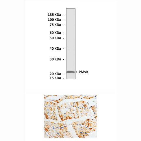
Product Sheet CA1067
Description
BACKGROUND Phosphomevalonate kinase (PMvK; EC 2.7.4.2.) catalyzes an essential step in the so-called mevalonate pathway, which appears to be the sole pathway for the biosynthesis of sterols and other isoprenoids in mammals and archea. Isoprenoid biosynthesis starts with three molecules of acetyl-CoA, which in a series of six different enzyme reactions are converted to isopentenyl pyrophosphate, the basic C5 isoprene unit used for the synthesis of all isoprenoids. PMvK catalyzes the fifth reaction of the pathway, which is the phosphorylation of phosphomevalonate to produce pyrophosphomevalonate. PMvK is a member of the GHMP1 kinase superfamily, which is composed of 13 families; eight of which have a known catalytic function. Superfamily members share a consensus sequence (PX 3GSSAA) that has been shown recently to form a unique P-loop structure. Among the features that distinguish the GHMP kinase nucleotide-binding pocket from the more well known P-loop kinases are that the GHMP P-loop is “missing” the catalytic lysine reside thought to stabilize negative charge that develops at the beta, gamma-bridging oxygen of ATP during bond cleavage, the nucleotide is positioned relative to the P-loop such that the adenosyl group occupies the pocket normally assigned to the gamma-phosphoryl acceptor, and the six-membered ring of the adenine is rotated into a position over the ribose (i.e. synrather than anti).1 PMvK is expressed in a variety of tissues with high expression in heart, skeletal muscle, liver, kidney, and pancreatic tissues.2 The human PMvK sequence contains a putative PTS-1, SRL, at the extreme carboxyl terminal. Analysis of PMvK message from lymphoblasts grown in lipoprotein-deficient media or media containing lovastatin, an inhibitor of HMG-CoA reductase, indicated that PMvK expression may be responsive to lipid availability. It was shown that PMvK is a peroxisomal protein which utilizes a PTS-1 for its peroxisomal localization.3 However other studies have also identified the chromosomal and cytosolic location of PMvK.3,4
REFERENCES
1. Pilloff, D. et al: J. Biol. Chem. 278:4510-5, 2002
2. Chambliss, K. L. et al: J. Biol. Chem. 271:17330-4, 1996
3. Olivier, L.M. et al: J. Lipid Res. 40:672-9, 1999
4. Hogenboom, M. et al: J. Lipd Res. 45:697-405, 2004
2. Chambliss, K. L. et al: J. Biol. Chem. 271:17330-4, 1996
3. Olivier, L.M. et al: J. Lipid Res. 40:672-9, 1999
4. Hogenboom, M. et al: J. Lipd Res. 45:697-405, 2004
Products are for research use only. They are not intended for human, animal, or diagnostic applications.
Details
Cat.No.: | CA1067 |
Antigen: | Short peptide from human PMvK sequence. |
Isotype: | Rabbit IgG |
Species & predicted species cross- reactivity ( ): | Human, Rat |
Applications & Suggested starting dilutions:* | WB 1:1000 IP n/d IHC 1:50 - 1:200 ICC n/d FACS n/d |
Predicted Molecular Weight of protein: | 22 kDa |
Specificity/Sensitivity: | Detects endogenous levels of PMvK proteins without cross-reactivity with other related proteins. |
Storage: | Store at -20°C, 4°C for frequent use. Avoid repeated freeze-thaw cycles. |
*Optimal working dilutions must be determined by end user.
Products
| Product | Size | CAT.# | Price | Quantity |
|---|---|---|---|---|
| Rabbit Phosphomevalonate Kinase Antibody: Rabbit Phosphomevalonate Kinase Antibody | Size: 100 ul | CAT.#: CA1067 | Price: $375.00 |

